Rare or complexed conditions

Misdiagnosed Conditions

Many rare or complexed conditions are sometimes misdiagnosed or have Doctors scratching their heads.

The world of illness is a MAZE that sometimes is worked out fast.

But put in a rare illness into the mix and this might take much longer to work out, and becomes a headache for all involved.

BUT you’re looking for the Doctor that thinks outside the box, as the saying goes.

Crohn’s disease is a condition where there is inflammation in the gut (gastrointestinal tract). The disease flares up from time to time. Symptoms vary, depending on the part of the gut affected and the severity of the condition. The most common symptoms are diarrhea, abdominal pain, and feeling generally unwell. Medication can often ease symptoms when they flare up. Surgery to remove sections of the gut is needed to treat some flare-ups. Medication taken regularly may prevent symptoms from flaring up.

Crohn’s disease is a condition that causes inflammation of the wall of the gut (gastrointestinal tract). Any part of the gut can be affected. This can lead to various symptoms (detailed below). Crohn’s disease is named after Dr. Crohn, the person who first described the disease in the 1930s.

What is inflammatory bowel disease?

When doctors talk of inflammatory bowel disease they usually mean people who either have Crohn’s disease or ulcerative colitis. Both these conditions can cause inflammation of the colon and rectum (large bowel or large intestine) with similar symptoms, such as bloody diarrhea, etc. Although these conditions are similar and treatments are similar, there are differences. For example, the inflammation of ulcerative colitis tends to be just in the inner lining of the gut (gastrointestinal tract), whereas the inflammation of Crohn’s disease can spread through the whole wall of the gut. Also, ulcerative colitis only affects the colon and rectum, whereas Crohn’s disease can affect any part of the gut.

However, about 1 in 20 people with inflammatory bowel disease affecting just the colon cannot be classified as having either Crohn’s disease or ulcerative colitis because they have some features of both conditions. This is sometimes called indeterminate colitis.

Note: inflammatory bowel disease is sometimes shortened to IBD. This is not the same as IBS which is short for irritable bowel syndrome – a very different disease.

The gut (gastrointestinal tract) is the long tube that starts at the mouth and ends at the anus. When we eat, food passes down the gullet (esophagus), into the stomach and then into the small intestine. The small intestine has three sections – the duodenum, jejunum, and ileum. In the small intestine, food is digested and absorbed into the bloodstream. The structure of the gut then changes to become the large intestine (colon and rectum, sometimes called the large bowel).

The colon absorbs water and contains food that has not been digested, such as fiber. This is passed into the last part of the large intestine, where it is stored as stools (feces). Feces are then passed out of the anus into the toilet.

Crohn’s disease is diagnosed in about 1 in 10,000 people every year. About 145 in every 100,000 people in the UK have Crohn’s disease. It can develop at any age but most commonly starts between the ages of 15 and 30. Crohn’s disease is more common in people who smoke. If you have a family member with Crohn’s disease, you are more likely to develop the condition yourself. It is also more common in people who have had their appendix removed, for the first five years after the operation.

In Crohn’s disease, one or more patches of inflammation develop in parts of the gut (gastrointestinal tract). Any part of the gut can be affected. However, the most common site for the disease first to start is the last part of the small intestine (the ileum). The ileum is affected in about half of cases. Other parts of the small intestine and the colon are also commonly affected. The mouth, gullet (oesophagus) and stomach are affected much less commonly.

A patch of inflammation may be small, or spread quite a distance along part of the gut. Several patches of inflammation may develop along the gut, with normal sections of gut in between. In about 3 in 10 cases, the inflammation occurs just in the small intestine. In about 2 in 10 cases the inflammation occurs just in the colon. In a number of cases, the inflammation occurs in different places in the gut.

The cause is not known. About 3 in 20 people with Crohn’s disease have a close relative who also has it. This means there may be some genetic factor. However, other factors such as a germ (bacterium or virus) may be involved. One theory is that a germ may trigger the immune system to cause inflammation in parts of the gut (gastrointestinal tract) in people who are genetically prone to develop the disease.

For more info

http://patient.info/health/crohns-disease-leaflet

http://www.mayoclinic.org/diseases-conditions/crohns-disease/basics/symptoms/con-20032061

YouTube Video Crohn’s disease

This animation describes the cause, symptoms, and factors in the development of Crohn’s disease.

Published on Oct 16, 2015

My Crohn’s Disease Story. What is Crohn’s Disease? What causes it? It’s a Inflammatory Bowel Disease IBD. Crohn’s is similar to Ulcerative Colitis. I’m just a teen struggling to live with this illness. I use the injection pen medicine Humira Adalimumab to treat my painful flare-ups. This is my story battling this condition. Visiting Doctors in the hospital for surgeries. You go to the bathroom a lot and are always sick. You always have to have a positive view on life.

SWAN stands for Syndrome Without A Name.

It is a term used to describe disabled children who are thought to have a genetic syndrome or condition that doctors have so far been unable to identify.  Many of these children have had lots of tests including blood tests, microarray, lumber punctures, EEG’s, ECG’s and MRI’s but they have all come back negative.

SWAN is not a condition, it is not a diagnosis and it does not refer to one specific syndrome or condition. In fact, the reason that many children remain undiagnosed is because it is highly likely that they have a very rare syndrome or condition.

Why is getting a diagnosis important?

Without a diagnosis life can be very isolating.  Families often feel like they don’t ‘fit in’ and without a diagnosis it can be very difficult to answer questions about their child.  They don’t know what the future holds for their child – Will they walk? Talk? What will their life expectancy be?

http://undiagnosed.org.uk/information-and-resources/what-does-swan-being-undiagnosed-mean

Undiagnosed – A Documentary

A documentary focussing on the struggles faced by families with children who have undiagnosed genetic conditions and the hope which research studies such as the Deciphering Developmental Disorders study provides them.

This documentary was independently produced by Kat Williams for her MA in Multimedia Broadcast Journalism.

http://undiagnosed.org.uk/undiagnosed-a-documentary

We have found a Mother who has set up a Facebook Group to help others dealing with very Rare Condition her own Daughter is one of Three Known

Cases WORLDWIDE

https://www.facebook.com/groups/1654366864833848/

Rare but manageable diagnosis PGS (posterior glottic stenosis) affecting 1 in 200,000 children. It is often caused by traumatic intubation and associated with prolonged intubation and easily misdiagnosed and often overlooked It is also accompanied with subglottic stenosis.
Background

Glottic stenosis is narrowing of the larynx at the level of the glottis (ie, vocal cords). It is caused by webbing, fibrosis, or scarring and most often involves the posterior glottis. The most common cause of stenosis is prolonged endotracheal intubation. In patients who are intubated for more than 10 days, the risk of developing posterior glottic stenosis is as high as 15%. Inflammation, infection trauma, and congenital and iatrogenic causes also contribute to glottic stenosis. In all cases, the preoperative evaluation must include direct laryngoscopy as well as microlaryngoscopy with an assessment of vocal cord mobility. Treatment is based on the etiology of the stenosis and the thickness of the stenotic segment. Various medical and surgical methods are discussed according to the type of stenosis.

Anatomical regions of the larynx are shown in the image below.

Congenital subglottic stenosis was first described by Rossi in 1826. Mackenzie in 1862 described laryngeal stenosis that results from inflammatory conditions, especially, syphilis which was predominant then. In the late 19th century, Von Schroetter performed laryngeal dilatation with rigid vulcanite tubes and used pewter plugs for stents.

In 1924, Haslinger described an endoscopic technique for web excision with the placement of a silver plate keel. In 1935, Iglauer described the use of a spring ring from a watch chain as a keel after glottic stenosis excision. In 1950, McNaught used a tantalum keel postglottic stenosis excision.

In 1979 and 1980, Dedo and Lichtenberger, respectively, modified this technique for clinical use. In 1980, Lichtenberger introduced a technique for the treatment of anterior commissure webs. This technique involved endoscopically suturing a keel in place from inside the airway out to the neck. In 1994, Lichtenberger reported on a series of 13 patients who were treated for anterior commissure webs with a keel-fixing technique that involved the use of a specialized endo-extralaryngeal needle carrier.

In 1968, Dedo and Sooy described an aryepiglottic fold mucosal flap. In 1984, they detailed the endoscopic use of the laser for mild posterior commissure stenosis and described the creation of a trapdoor flap.In 1973, Montgomery recommended a superiorly based advancement mucosal flap from the interarytenoid area. Multiple variations of the laryngofissure approach have been developed.

In 1993, Zalzal described an anterior laryngofissure technique with posterior cricoidotomy and cartilage grafting. In 1995, Biavati et al successfully used, in 5 children, a single-stage procedure for the repair of congenital laryngeal webs that were associated with subglottic stenosis. Recent work has centered around endoscopic techniques for repair, use of a carbon dioxide laser or microdebrider for excision of webs, topical application of mitomycin-C, and, lately, chitosan for prevention of restenosis.

Problem

Glottic stenosis may accompany subglottic stenosis or may be diagnosed as a separate entity. Glottic stenosis may be a congenital or an acquired condition, represented as anterior or posterior webs, interarytenoid adhesions with or without impaired vocal cord mobility, or complete fusion of the true vocal folds (rare).

Epidemiology

Frequency

Acquired posterior glottic stenosis is the most common form of glottic stenosis and typically results from trauma due to endotracheal intubation. The risk of developing posterior glottic stenosis is reported to be as high as 15% in patients who are intubated for more than 10 days. Factors that contribute to increased risk of stenosis related to intubation include traumatic intubation, prolonged duration of intubation, multiple extubations and reintubations, an oversized endotracheal tube used for intubation, motion of the patient or the endotracheal tube, gastroesophageal reflux, and local infection.

Congenital glottic stenosis is a rare disorder and may exist as a thin membranous stenosis, a thick anterior or posterior web, or as a complete fusion of the vocal cords. Congenital laryngeal webs are rare; the largest study identified 51 children over a 32-year period. In 2009, Cheng et al reported on 4 children with Shprintzen syndrome who had a severe congenital anterior glottic web.Crowe et al have reported on a rare case of glottic stenosis in an infant with Fraser syndrome.

For more on this click on the link below.

http://emedicine.medscape.com/article/864439-overview#a8

With this site we hope to create more awareness for the rare congenital anomaly of Laryngeal Cleft. Besides that, we hope to share experiences and information with other parents and between them. Get in contact with other parents now!

What is laryngeal cleft?
Laryngeal cleft (fissure of the larynx) is a rare abnormality of the separation between the larynx, or voice box, and the esophagus. When the larynx develops normally it is completely separate from the esophagus, so swallowed foods go directly into the stomach. A laryngeal cleft creates an opening between the larynx and the esophagus so food and liquid can pass through the larynx into the lungs.

What are the symptoms of laryngeal cleft?
Laryngeal cleft causes swallowing problems. Coughing, gagging, frequent respiratory infections, and chronic lung disease are also symptoms of the disorder.

How is laryngeal cleft diagnosed?
Laryngeal cleft is diagnosed through a comprehensive aero-digestive evaluation performed under anesthesia.

What is the treatment for laryngeal cleft?
Laryngeal cleft is treated with surgery to close the opening in the larynx. Conventional treatment for the condition is invasive surgery requiring an incision in the neck and opening of the larynx. Surgeons at Children’s Hospital Boston were the first in the world to perform robotic surgery on an airway to correct a laryngeal cleft. The robotic equipment allows surgeons to work through the oral cavity and in the restricted confines of the airway without impeding breathing. This minimally-invasive technique results in less pain and scarring and faster recovery. At this time we are constructing this website and development will be ongoing.

http://www.laryngeal-cleft.com/What-is-laryngeal-cleft

What is Hirschsprung disease?

Hirschsprung disease is an intestinal disorder characterized by the absence of nerves in parts of the intestine. This condition occurs when the nerves in the intestine (enteric nerves) do not form properly during development before birth (embryonic development). This condition is usually identified in the first two months of life, although less severe cases may be diagnosed later in childhood.

Enteric nerves trigger the muscle contractions that move stool through the intestine. Without these nerves in parts of the intestine, the material cannot be pushed through, causing severe constipation or complete blockage of the intestine in people with Hirschsprung disease. Other signs and symptoms of this condition include vomiting, abdominal pain or swelling, diarrhea, poor feeding, malnutrition, and slow growth. People with this disorder are at risk of developing more serious conditions such as inflammation of the intestine (enterocolitis) or a hole in the wall of the intestine (intestinal perforation), which can cause serious infection and may be fatal.

There are two main types of Hirschsprung disease, known as short-segment disease and long-segment disease, which are defined by the region of the intestine lacking nerve cells. In short-segment disease, nerve cells are missing from only the last segment of the large intestine. This type is most common, occurring in approximately 80 percent of people with Hirschsprung disease. For unknown reasons, the short-segment disease is four times more common in men than in women. Long-segment disease occurs when nerve cells are missing from most of the large intestine and is the more severe type. Long-segment disease is found in approximately 20 percent of people with Hirschsprung disease and affects men and women equally. Very rarely, nerve cells are missing from the entire large intestine and sometimes part of the small intestine (total colonic aganglionosis) or from all of the large and small intestine (total intestinal aganglionosis).

Hirschsprung disease can occur in combination with other conditions, such as Waardenburg syndrome, type IV; Mowat-Wilson syndrome; or congenital central hypoventilation syndrome. These cases are described as syndromic.

Hirschsprung disease occurs in approximately 1 in 5,000 newborns.

What genes are related to Hirschsprung disease?

Isolated Hirschsprung disease can result from mutations in one of several genes, including the RET, EDNRB, and EDN3 genes. However, the genetics of this condition appear complex and are not completely understood. While a mutation in a single gene sometimes causes the condition, mutations in multiple genes may be required in some cases. The genetic cause of the condition is unknown in approximately half of affected individuals.

http://ghr.nlm.nih.gov/condition/hirschsprung-disease

Understanding Hirschsprung’s Disease
A guide for parents and carers
link

Achalasia Overview

Achalasia is an uncommon swallowing disorder that affects about 1 in every 100,000 people. The major symptom of achalasia is usually difficulty with swallowing. Most people are diagnosed between the ages of 25 and 60 years. Although the condition cannot be cured, the symptoms can usually be controlled with treatment.

Achalasia Cause

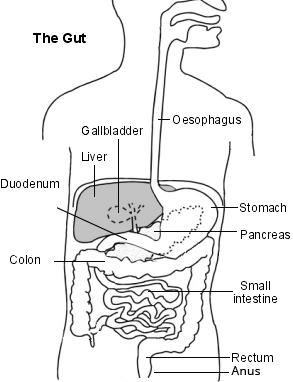
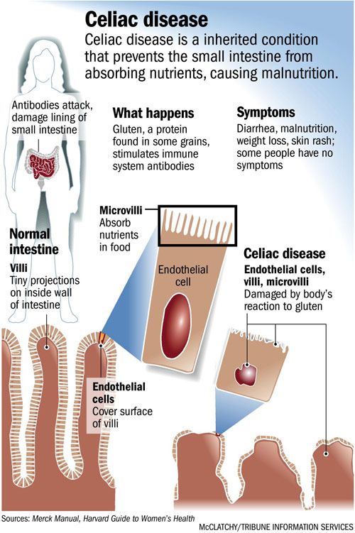
In achalasia, nerve cells in the esophagus (the tube that carries food from the mouth to the stomach) degenerate for reasons that are not known. The loss of nerve cells in the esophagus causes two major problems that interfere with swallowing.

1/ The muscles that line the esophagus do not contract normally, so that swallowed food is not propelled through the esophagus and into the stomach properly.

2/ The lower esophageal sphincter (LES), a band of muscle that encircles the lower portion of the esophagus, does not function correctly.

3/ Normally, the LES relaxes when we swallow to allow swallowed food to enter the stomach. When the food has moved through the esophagus into the stomach, the LES muscle contracts to squeeze the end of the esophagus closed, thus preventing the stomach contents from flowing backward (refluxing) into the esophagus.

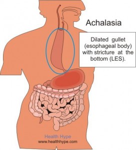
In people with achalasia, the LES fails to relax normally with swallowing. Instead, the LES muscle continues to squeeze the end of the esophagus, creating a barrier that prevents food and liquids from passing into the stomach. Over time, the esophagus above the persistently contracted LES dilates, and large volumes of food and saliva can accumulate in the dilated esophagus.

Achalasia Symptoms

The most common symptom of achalasia is difficulty swallowing. Patients often experience the sensation that swallowed material, both solids and liquids, gets stuck in the chest. This problem often begins slowly and progresses gradually. Many people do not seek help until symptoms are advanced. Some people compensate by eating more slowly and by using maneuvers, such as lifting the neck or throwing the shoulders back, to improve emptying of the esophagus.

Other symptoms can include chest pain, regurgitation of swallowed food and liquid, heartburn, difficulty burping, a sensation of fullness or a lump in the throat, hiccups, and weight loss.

Achalasia Diagnosis

Achalasia may be suspected based upon symptoms, but tests are needed to confirm the diagnosis.

Chest x-rays — A chest x-ray may reveal a dilated esophagus and absence of air in the stomach. However, a chest x-ray is not adequate for a diagnosis of achalasia and further testing is required.

Barium swallow test — The barium swallow test is a common screening test for achalasia. The test involves swallowing a chalky-tasting, thick mixture of barium while x-rays are taken. The barium shows the outline of the esophagus and lower esophageal sphincter (LES). Characteristic findings of achalasia on barium swallow include a persistently narrowed region at the end of the esophagus (the LES), with a dilated esophagus above the narrowed region. The barium swallow may also show spastic contractions in the esophagus above the LES, a condition called “vigorous achalasia”.

Esophageal manometry (also called esophageal motility study) — Manometry is a test that measures changes in pressures within the esophagus that are caused by the contraction of the muscles that line the esophagus. The test involves the passage of a thin tube through the mouth or nose into the esophagus. The tube is lined by numerous pressure sensors that convey pressures within the esophagus to a device that records those pressures. Patients are usually instructed to have nothing to eat or drink for eight hours before the test, and they are given sips of water to swallow while the tube is in place.

Manometry is almost always used to confirm the diagnosis of achalasia. The test typically reveals three abnormalities in people with achalasia: high pressure in the LES at rest, failure of the LES to relax after swallowing, and an absence of useful (peristaltic) contractions in the lower esophagus. The last two features are the most important and are required to make the diagnosis of achalasia.

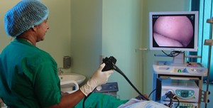
Endoscopy — Endoscopy allows the physician to see the inside of the esophagus, LES, and stomach using a thin, lighted, flexible tube. Most patients are given sedatives during the endoscopy procedure. This test is usually recommended for people with suspected achalasia and is especially useful for detecting other conditions that can mimic achalasia such as cancer of the upper portion of the stomach.

http://www.uptodate.com/contents/achalasia-beyond-the-basics

Towards a global classification for mast cell disorders

Keywords: Mast cells, tryptase, histamine, mastocytosis, allergy, anaphylaxis, urticaria

The term “mast cell activation syndrome (MCAS)” is finding increasing use as a diagnosis for individuals who present with signs and symptoms involving the dermis, gastrointestinal track and cardiovascular system; frequently accompanied by neurologic complaints. Such patients often have undergone multiple extensive medical evaluations by different physicians in varied disciplines without a definitive medical diagnosis until the diagnosis of “MCAS” is applied. However, “MCAS” as a distinct clinical entity has not been generally accepted nor do there exist definitive criteria for diagnosis. Based on current understanding of this disease “syndrome” and on what we do know about mast cell activation and resulting pathology, we will explore and propose criteria for its diagnosis. The proposed criteria will be discussed in the context of other disorders involving mast cells or with similar presentations; and as a basis for further scientific study and validation.

The last several years have witnessed an increasing use of the term “mast cell activation syndrome (MCAS)” as a diagnosis for individuals who present with signs and symptoms from flushing to hives, abdominal pain to diarrhea, and paresthesias to cognitive dysfunction Repository for an example of what is available to the lay public, and where an extensive medical evaluation has failed to identify an etiology. Although “MCAS” as a distinct clinical entity has not been yet recognized nor defined by definitive criteria for diagnosis, there have been scientific publications on the subject, even relating to a possible genetic basis of a “mast cell mediator syndrome” This article will explore the evolution of the application of the term “MCAS” and propose criteria for its diagnosis in the context of other disorders involving mast cells or with similar presentations; and as a basis for further scientific study and validation.

http://www.ncbi.nlm.nih.gov/pmc/articles/PMC3753019/#SD1

Sandifer syndrome misdiagnosed as refractory partial seizures in an adult.

We report a 27-year-old man with an intellectual disability and no prior history of seizures who presented with episodes of abdominal pain, head/eye version, and unresponsiveness that were misdiagnosed and treated as partial seizures. Associated vomiting and haematemesis led to the correct diagnosis and treatment of reflux oesophagitis. The episodes immediately resolved and a diagnosis of Sandifer syndrome was made. This is only the second report of Sandifer syndrome in adult, a movement disorder of unknown mechanism that occurs almost exclusively in young children, often misdiagnosed as epilepsy or episodic dystonia.

http://www.ncbi.nlm.nih.gov/pubmed/15075069

The neurological features of Sandifer syndrome usually involve spasmodic torticollis, or chronic involuntary movements of the neck in right, left, up or down directions, and dystonia, in which sustained muscle contractions cause twisting and writhing movements and abnormal postures. Although this condition does not have a neurological basis, it is often misdiagnosed as epileptic seizures due to these neurological signs. The spasms associated with this syndrome may last several minutes and can occur frequently throughout the day, although they are often associated with feeding.

Sandifer’s Syndrome: a Misdiagnosed and Mysterious Disorder

Sandifer’s Syndrome (SS) is a rare pediatric manifestation of gastroesophageal reflux (GER) disease characterized by abnormal and dystonic movements of the head, neck, eyes, and trunk. Although Sandifer initially observed the association, Kinsbourne and Oxon first reported it based on the observations of Sandifer. The syndrome is most certainly underrecognized, and delays in diagnosis are due to atypical presentations or cases in which the diagnosis is not part of the differential.

A 9-month-old boy was referred with a 2 months history of vomiting. 15 days ago before admitting to our hospital, he started to make bizarre head and neck movements as told by his parents. The parents provided careful video recording of these movements. The most striking feature pronounced neck dystonia with a repeated rotation of the neck and tilting of the head towards the left shoulder. These movements were observed during or just after the feeding. Sometimes upward deviation of the eyes and head nodding accompanied these movements. All of these movements stopped when he was asleep. The milestones of motor and mental development were normal. His physical and neurological examinations were normal.

http://www.ncbi.nlm.nih.gov/pmc/articles/PMC4025139/

Celiac disease is an immune reaction to eating gluten, a protein found in wheat, barley, and rye. If you have celiac disease, eating gluten triggers an immune response in your small intestine. Over time, this reaction produces inflammation that damages the small intestine’s lining and prevents absorption of some nutrients (malabsorption).

The intestinal damage can cause weight loss, bloating and sometimes diarrhea. Eventually, your brain, nervous system, bones, liver and other organs can be deprived of vital nourishment. In children, malabsorption can affect growth and development. The intestinal irritation can cause stomach pain, especially after eating. There’s no cure for celiac disease — but following a strict gluten-free diet can help manage symptoms and promote intestinal healing. Your small intestine is lined with tiny hair-like projections called villi, which work to absorb vitamins, minerals and other nutrients from the food you eat. Celiac disease damages the villi, leaving your body unable to absorb nutrients necessary for health and growth.

10 Symptoms of Celiac Disease: Do You Have It?

Celiac disease, also known as coeliac disease, is an autoimmune disorder that affects around 1 in 100 Americans. The disease is caused by gluten intolerance. Gliadin, a protein in wheat, causes the afflicted person’s immune system to attack its own bowel tissue. This causes villous atrophy or the erosion of the nutrient-absorbing villi that line the small intestine. Other grains, like barley and rye, have similarly shaped proteins which can cause the same reaction in sensitive individuals.

Celiac disease can cause irreversible damage. The issue seems to be genetic in cause and can be passed down through generations. Testing for celiac disease is done through blood tests and endoscopy. It is important for the afflicted person to continue eating gluten while they are tested for celiac disease. When they remove gluten from their diet, their body starts to repair itself, giving a false negative for the tests.

Celiac disease can cause a multitude of problems. The major ones come from malabsorption of nutrients. This can lead to osteoporosis, anemia, and stunted growth in children. Those who suffer from celiac disease are also more likely to develop non-Hodgkin lymphoma when compared to the general population. The best way to protect yourself and minimize your risk is early diagnosis and strict diet.

Here are 10 symptoms of celiac disease. If you suffer from any of these symptoms or believe you may be at risk for celiac disease, please contact your doctor for testing.

1. No Symptoms

Unfortunately, many people who suffer from celiac disease have no symptoms. This is particularly dangerous as intestinal damage can occur even when no symptoms are present. With new screening techniques, people are being diagnosed without ever suspecting they have the disease. It is important for people with no symptoms but a positive diagnosis to eat properly, as they are still at risk for serious complications of celiac disease, such as cancer.

2. Bloating

Many people who have celiac disease suffer from painful bloating. For days after consuming gluten, the stomach may be distended and filled with gas. This can be quite painful. Many people complain of intestinal pain and the feeling of being “6 months pregnant”. They may also suffer from excessive gas and flatulence. Bloating can be symptoms of other dietary conditions, so look for it in conjunction with the consumption of gluten.

3. Diarrhea

Chronic diarrhea is a common symptom of those afflicted with celiac disease. The episodes may be smelly, explosive, and painful. The color of the stool may be pale or white, and typically floats on the surface of the water. Even solid stools may present these conditions. Related symptoms to chronic diarrhea include haemorrhoids. These can occur when there is not enough substance in the stools.

4. Constipation

While diarrhea is common in celiac sufferers, some people may be afflicted with constipation. This is from the body sluggishly moving the contents of the bowels. The body may be trying to pull all the nutrients possible from the food as the damaged villi will not work as efficiently. The constipation may be marked by periods of heavy diarrhea as the person consumes gluten. The constipation may cause pain and bloating.

5. Weight Loss or Gain

Many people who suffer from celiac disease lose weight as their body reacts to the missing nutrients. The diarrhea caused by gluten may stall the absorption of calories from the food, purging the body from its fuel. Conversely, a sufferer may in fact gain weight instead of lose it. Their bodies may be screaming for nutrients, so they eat in excess to achieve proper nutrition. They may have a hard time losing weight until they restrict gluten from their diet.

6, 7, 8, 9, 10, pages

http://www.activebeat.co/health-news/10-symptoms-of-celiac-disease-do-you-have-it-2/6/

This is a Rare Condition

Many thanks to Doctors Melissa Palmer and Emil Miskovski for their help in writing this article.

Primary Biliary Cirrhosis (PBC) is a chronic liver disease that slowly destroys the bile ducts within the liver (intrahepatic bile ducts). Liver inflammation over a period of years may cause scarring which leads to cirrhosis. PBC is NOT alcohol or drug related, and it is NOT contagious.

The name “Primary Biliary Cirrhosis” is somewhat deceiving since cirrhosis only occurs in the last stage of the disease (stage 4) after many years of inflammation. With early diagnosis and proper medications, most with PBC will never reach the cirrhosis stage of PBC.

PBC is also called “Chronic Nonsuppurative Destructive Cholangitis” and “Primary Autoimmune Cholangitis.” However, these alternative terms are not widely accepted and therefore not in use.

Definition from New American Medical Dictionary:
Primary: First in order of development, most important, arising spontaneously.
Biliary: Relating to or affecting the bile duct system or bile.
Cirrhosis: An inflammatory disease of the liver associated with the replacement of liver cells by fibrous tissue. Passage of blood through the liver may eventually be obstructed by the cirrhosis.


The cause of PBC is still unknown, but it is not alcohol or drug induced. Current studies suggest it may involve autoimmunity, infection, or genetic predisposition, and does seem to appear more often in certain families. Women are affected 10 times more than men, and PBC is usually diagnosed in patients between the ages of 35 to 60 years.

Those with PBC usually look extremely healthy, and many are 10 to 30 pounds overweight. The slight bronze pigmentation of the skin is often present in the advanced stage of the disease, and makes the individual look tanned. The outward appearances doesn’t tell the story of what is going on inside their bodies. Even on the transplant list stage, many with PBC look healthy. A person with PBC commonly hears comments such as “you look so healthy or you don’t look sick.”

There is a lot on this from the link below

http://pbcers.org/about-pbc/

Hydrocephalus comes from the Greek hydro meaning water and cephalie, meaning brain. A watery fluid, known as cerebro-spinal fluid or CSF, is produced constantly inside each of the four spaces or ventricles inside the brain: between 400 and 600mls is produced each day. The CSF normally flows through narrow pathways from one ventricle to the next, then out over the outside of the brain and down the spinal cord. The CSF is absorbed into the bloodstream, and the amount and pressure are normally kept within a fairly narrow range.

If the drainage of CSF is prevented at any point, the fluid accumulates in the ventricles inside the brain, causing them to swell and resulting in compression of the surrounding tissue. In babies and infants, the head will enlarge. In older children and adults, the head size cannot increase as the bones which form the skull are completely joined together.

What causes hydrocephalus?

The condition is caused by the inability of CSF to drain away into the bloodstream. There are many reasons why this can happen. Hydrocephalus can be congenital or acquired.

Key

Cerebrum: The main part of the brain, in two halves one on each side (left and right hemispheres).

Cerebellum: The smaller part of the brain at the back of the head, involved in muscular coordination.

Ventricles: Connected spaces inside the brain, where CSF is produced. There are two lateral ventricles (one each side), a third ventricle and a fourth ventricle. CSF flows from here over the outside of the brain before being absorbed back into the bloodstream.

Choroid plexus: The organ that makes CSF.

Superior sagittal sinus: The major vein that runs over the centre of the top of the brain and collects CSF, draining it back into the central bloodstream.

Pituitary gland: A gland situated in the brain cavity but just behind the face. It is very important in secreting a large number of hormones that regulate body function and development. An example is growth hormone, and others are involved in sexual development.

Brainstem: A very important area connecting the spinal cord to the brain. It controls breathing and other vital functions.

Spinal cord: A complex trunk of nerves transmitting impulses to all parts of the body, allowing muscle movement, sensation and reflexes.

http://www.shinecharity.org.uk/hydrocephalus

http://www.nhs.uk/conditions/Hydrocephalus/Pages/Introduction.aspx

http://www.brainandspine.org.uk/hydrocephalus-water-brain-and-shunts?gclid=CI7ctIqR9coCFUEaGwodhxMKSQ

Mitochondrial diseases result from failures of the mitochondria, specialized compartments present in every cell of the body except red blood cells. Mitochondria are responsible for creating more than 90% of the energy needed by the body to sustain life and support growth. When they fail, less and less energy is generated within the cell. Cell injury and even cell death follow. If this process is repeated throughout the body, whole systems begin to fail, and the life of the person in whom this is happening is severely compromised. The disease primarily affects children, but adult onset is becoming more and more common.

Diseases of the mitochondria appear to cause the most damage to cells of the brain, heart, liver, skeletal muscles, kidney and the endocrine and respiratory systems.

Depending on which cells are affected, symptoms may include loss of motor control, muscle weakness and pain, gastro-intestinal disorders and swallowing difficulties, poor growth, cardiac disease, liver disease, diabetes, respiratory complications, seizures, visual/hearing problems, lactic acidosis, developmental delays and susceptibility to infection.

More info below

http://www.umdf.org/site/pp.aspx?c=8qKOJ0MvF7LUG&b=7934627

https://ghr.nlm.nih.gov/search?query=Mitochondrial+Disease

MRC Centre for Translational Research in Neuromuscular Disease Mitochondrial Disease Patient Cohort (UK)

Mitochondria are found in every cell in the human body and function like tiny batteries, converting energy locked in food into a form that can be used by the cell. These tiny batteries contain their own genetic information (mtDNA) that is passed from mother to child, and acts as a blueprint for parts of the enzymes crucial for the energy conversion process. Genes stored in the nucleus of the cell are also important for normal mitochondrial function. Disruption of either mtDNA or nuclear DNA (nDNA) can cause the batteries to fail, resulting in mitochondrial disease.

Mitochondrial disease affects at least 1 in 6500 people in the UK and commonly leads to symptoms of fatigue, weakness, unsteadiness,swallowing difficulties, stroke-like episodes, seizures, acidosis and heart disease. Recognition of mitochondrial disease is a significant clinical problem and many of the different forms of the disease are still being characterized as virtually any body organ can be affected. At present no cure is available and symptoms are treated with conventional medicines that have not been assessed in patients specifically with mitochondrial disease.

 Mitochondria are found in every cell in the human body and function like tiny batteries, converting energy locked in food into a form that can be used by the cell. These tiny batteries contain their own genetic information (mtDNA) that is passed from mother to child, and acts as a blueprint for parts of the enzymes crucial for the energy conversion process. Genes stored in the nucleus of the cell are also important for normal mitochondrial function. Disruption of either mtDNA or nuclear DNA (nDNA) can cause the batteries to fail, resulting in mitochondrial disease.

Mitochondrial disease affects at least 1 in 6500 people in the UK and commonly leads to symptoms of fatigue, weakness, unsteadiness, swallowing difficulties, stroke-like episodes, seizures, acidosis and heart disease. Recognition of mitochondrial disease is a significant clinical problem and many of the different forms of the disease are still being characterized as virtually any body organ can be affected. At present no cure is available and symptoms are treated with conventional medicines that have not been assessed in patients specifically with mitochondrial disease.

The Mitochondrial Disease Patient Cohort will define a cohort of 1000 patients, (adults and children), in whom mitochondrial disease has been identified clinically and/or genetically. Our aims in developing this cohort are to translate improvements in our understanding of the science of mitochondrial disease into direct health benefits for patients. We also want to further our understanding of mitochondrial disease mechanisms and transmission and to evaluate (and optimise) the treatment of complications such as stroke, seizures, diabetes, poor growth and heart disease in patients with mitochondrial disease. The cohort will also be a rapidly accessible resource for assessing novel clinical interventions such as drugs targeted to mitochondria and exercise therapy.

The cohort is managed through the Medical Research Council (MRC) Centre for Translational Research in Neuromuscular Disease. Data collected will be managed and stored responsibly and patient care will be unaffected by their decision to enrol in the cohort or not. Patients are required to sign a consent form prior to their anonymised data being entered on to the cohort.

For further information on the MitoCohort, or to be part of it, please email: mitonews@ncl.ac.uk.

http://mitocohort.ncl.ac.uk/