Both very challenging conditions for those caring for Someone with these.

vacterl – charge

The VACTERL association refers to a recognized group of birth defects which tend to have a non-random occurrence. Note that this pattern is a recognized association, as opposed to a syndrome, because there is no known pathogenetic cause to explain the grouped. CHARGE syndrome was formerly referred to as CHARGE association, which indicates a non-random pattern of congenital anomalies that occurs together more frequently than one would expect on the basis of chance. Very few people with CHARGE will have 100% of its known features.


Vacterl-Charge

This website was written by a Parent who did not understand the medical talk, like the rest of us no doubt, so decided to do something about it, and in doing so, helps the rest of the Parents looking to understand better. When you think about scoliosis, the first thing that comes to mind is idiopathic scoliosis (which just means that the spine curves for no known reason, with no changes in the actual bones of the spine).  This is the typical kind of scoliosis you hear of when children get checked at the doctor or school by bending over to see if their back is straight. However, there is a rarer subset of scoliosis called Congenital scoliosis, then there is something called Hemivertebrae are usually wedge-shaped vertebrae, in between other vertebrae.  There are three main types of hemivertebrae, this website deals with understanding better what your Doctors are talking about.

it talks about

 Congenital scoliosis

Treatment for Congenital Scoliosis

Hemivertebrae – hemi·ver·te·bra/ (hem″e-ver´tĭ-brah

Butterfly Vertebrae

Block Vertebrae

Congenital Bar

Links for more information

http://conni60640-ivil.tripod.com/congenitalscoliosis101/index.html

ALSO

There is a Support group for this.

This group is for those dealing with scoliosis from malformations in the bones of the spine such as hemivertebra, butterfly vertebra, congenital bars, etc. Congenital scoliosis is fairly rare and accounts for about 15% of all cases of scoliosis and the treatment options can be different from those for idiopathic scoliosis (a curve for no known reason).

https://www.facebook.com/groups/congenitalscolisupport/

Vacterl-Charge

When our third child Matthew was born with a rare genetic disorder it only changed what was important in life…that is, everything is a gift and it is up to us how we respond to what is presented to us in life. After overcoming the adjustments of life with a special needs child, we decided we were not done with having children. God has chosen to bless us with another child with the same genetic disorder even though it is almost statistically impossible. Isaac is now a part of our continuing story of faith, perseverance and joy.

I started sharing stories of our journey over five years ago in a blog title Lessons from Matthew. At first I thought sharing our story was a creative expression and a way to help others. What I have found it that it helps me just as much. It helps my wife and the rest of our family reach out and see people, hurting people more clearly and it helps to put our own struggles into perspective. In return, we are strengthened. We have become a voice of hope and encouragement as we talk and interact with doctors, nurses, caregivers and to the groups we have been asked to speak and share our story with. We have spent countless hours in surgery waiting rooms, much more time, measured in days and months in hospital rooms and immeasurably more hours with caregivers and nurses in our home over the past 14 years sharing our faith in words, but even more importantly in our actions and in serving through a listening ear and through loving people right where they are at. As it often turns out, we have been blessed just as much as Matthew and Isaac have touched many lives and have been embraced by thousands as they have laughed with us, cried with us and prayed us through each step of their lives.

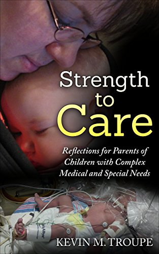
Strength to Care – Reflections for Parents of Children with Complex Medical and Special Needs is a collection of stories, or lessons, that all share a common theme of a hospital stay, surgery, complication or just plain old life is really hard right now. It is our prayer that our journey encourages you and gives you strength to care more deeply and with more understanding as you take care of the ones who have been placed in your care.

Vacterl-Charge

27-10-2017

Abstract

Background: Traditional teaching states that if the base of a sacral dimple is visible, associated spinal cord abnormalities are unlikely. In our experience, we noted that in several patients this was not true. We, therefore, set out to evaluate the incidence of spinal cord abnormalities in a cohort of children with a sacral dimple.

Objective: Our objective was to determine the frequency of spinal cord abnormalities detected on whole-spine MRI in children with a pure sacral dimple (cutaneous dimple without associated local pigmentation or hairy patch). The frequency of neurosurgical intervention in those with a spinal cord malformation was also calculated.

Materials and methods: We retrospectively reviewed all patients under the age of 16 years with clinically confirmed pure sacral dimple, who had undergone spinal MRI between 2005 and 2016. Patients with coccygeal pits were excluded from the study. We analyzed the MRI findings, calculated the incidence of cord abnormalities, and proportion of those who subsequently underwent neurosurgical procedures, such as spinal cord untethering. Other associated abnormalities were documented.

Results: During the 11-year study period, 33 patients (19 boys) with confirmed sacral dimple and whole-spine MRI were identified. The median age at the time of MRI was 21 months (range 2 months to 13 years). Eleven patients (33.3%) had abnormal cord detected on the MRI. Out of them, 4 had tethered cord, 2 had low lying cord (L2/3 level). Five had lesions related to the filum terminale (dermoid, cyst, lipoma). Of the four patients with tethered spinal cord, 2 had untethering procedures, 2 so far have been managed conservatively. Of the 7 patients with other cord abnormalities, 1 had excision of sacral dermoid. Nine of the 33 patients (27%) had other congenital anomalies, including 5 with syndromes.

Conclusion: In our experience, the incidence of spinal cord abnormalities in children with pure sacral dimple was 33.3%, some of which required surgery. Other congenital abnormalities were common. We recommend whole spinel MRI and a careful search for other congenital abnormalities.

http://www.jpss.eu/index.php/component/k2/item/927

Genomic microarray

Genomic microarray (Array-CGH) is an advanced technique in genetic testing that detects copy number changes in a person’s chromosomes. This means it looks for where there are deletions (bits missing) or duplications (where there are extra bits) in your DNA that would not be identified using conventional microscopy-based chromosome analysis (Karyotype). Such tests are now becoming more widely available in the NHS.

Many children with developmental delay who have had a ‘normal’ result from a microscopy-based chromosome analysis (Karyotype) in the past have, after consultation with their parents, been retested using genomic microarray analysis. A number of these children have been found to have a microdeletion or microduplication, therefore, offering the family an explanation for their child’s problems.

http://undiagnosed.org.uk/information-and-resources/how-can-i-get-a-diagnosis-for-my-child

22q11.2 deletion syndrome

DiGeorge syndrome, also called 22q11.2 deletion syndrome, is a disorder caused by a defect in chromosome 22. It results in the poor development of several body systems. Medical problems commonly associated with DiGeorge syndrome include heart defects, poor immune system function, a cleft palate, complications related to low levels of calcium in the blood, and delayed development with behavioral and emotional problems.

 The number and severity of symptoms associated with DiGeorge syndrome vary greatly. However, almost everyone with DiGeorge syndrome needs treatment from specialists in a variety of fields.

Before the discovery of the chromosome 22 defect, the disorder was known by several names — DiGeorge syndrome, velocardiofacial syndrome, Shprintzen syndrome, CATCH22 and others. Although the term “22q11.2 deletion syndrome” is frequently used today — and is generally a more accurate description — previous names for the disorder are still used.

Signs and symptoms of DiGeorge syndrome can vary significantly in type and severity, depending on what body systems are affected and how severe the defects are. Some signs and symptoms may be apparent at birth, but others may not appear until later in infancy or early childhood.

 Signs and symptoms may include some combination of the following:

http://www.mayoclinic.org/diseases-conditions/digeorge-syndrome/basics/symptoms/con-20031464

DiGeorge Syndrome (22q11.2 Deletion Syndrome, Velocardiofacial Syndrome)

http://genetics4medics.com/digeorge-syndrome.html

Digeorge syndrome – YouTube Video very interesting


Esophageal atresia is a common and life-threatening birth defect with a poorly understood etiology. In this study, we analyzed the sequence variants of coding regions for a set of esophageal atresia-related genes including MYCNSOX2CHD7GLI3, FGFR2 and PTEN for mutations using PCR-based target enrichment and next-generation sequencing in 27 patients with esophageal atresia. Genomic copy number variation analysis was performed using Affymetrix SNP 6.0. We found a de novo heterozygous mutation in the N-terminal region of the GLI3 gene (c.332 T > C, p.M111T) in a patient with esophageal atresia and hemivertebrae. The N-terminal region (amino acids 1–397) of GLI3 contains the repressor domain, which interacts with SKI family proteins. Using the co-immunoprecipitation assay, we found that interaction of GLI3 with the SKI family protein SKIL was significantly compromised by the p.M111T mutation ofGLI3. Thus far, all the identified mutations mapped within the repressor domain of GLI3were nonsense and frame-shift mutations. In this study, a missense mutation was initially detected in this region. Our finding is the first to link this GLI3 gene mutation with esophageal atresia in humans, which was previously suggested in an animal model.

Esophageal atresia (EA) is a developmental defect of the upper gastrointestinal tract in which the continuity between the upper and lower esophagus is lost. Esophageal atresia with or without tracheoesophageal fistula (TEF) is a rare malformation, occurring in approximately 1 in 3500 births. Although the underlying molecular mechanism remains obscure in most patients with EA/TEF, it has been associated with a wide spectrum of genetic syndromes caused by genetic alterations including mutations in single genes or chromosome aberrations.

Approximately 6% to 10% of syndromic EA/TEF is associated with chromosomal anomalies. The majority is represented by trisomies (trisomy 13, 18, 21) and or microdeletions of 22q11, 17q22–17q23.3 and 16q24.1. Meanwhile, mutations of MYCNSOX2 and CDH7 have also been reported to be responsible for syndromic EA, such as Feingold syndromeAEG syndrome (Anophthalmia-esophageal–genital syndrome) and CHARGE association (coloboma, heart anomalychoanal atresiaretardation, and genital and ear anomalies). Deletions and mutations in the GLI3 andFGFR2 genes are responsible for Pallister–Hall syndrome and Apert syndrome, respectively, in which EA is infrequently reported and  in rare cases, mutations in PTEN have been reported in the VATER association(vertebral defects, anal atresia, tracheoesophageal fistula with esophageal atresia, and radial or renal dysplasia).

In this study, we searched for sequence variants in the protein-coding regions of the human MYCNSOX2CHD7GLI3FGFR2 and PTEN genes by PCR-based target enrichment followed by next-generation sequencing using the Ion Torrent Personal Genome Machine™ (PGMTM, Life Technologies, Carlsbad, CA). We also analyzed genomic copy number variants (CNVs) in 27 patients with EA/TEF. We found a de novoheterozygous mutation in the N-terminal region of the GLI3 gene (c.332 T > C, p.M111T). Our finding is the first to link the GLI3 gene mutation with esophageal atresia in humans, which was previously suggested in an animal model.

CHARGE and VACTERL

Find on this page

Feingold Syndrome ALSO found on this website

Also found on this page

https://ghr.nlm.nih.gov/condition/feingold-syndrome

AEG syndrome

http://disorders.eyes.arizona.edu/category/alternate-names/aeg-syndrome

Anophthalmia-esophageal–genital syndrome

https://search.yahoo.com/search?p=(anophthalmia-esophageal%E2%80%93genital+s%C3%ADndrome&fr=yset_chr_syc_oracle&type=orcl_default

Choanal Atresia

http://www.nytimes.com/health/guides/disease/choanal-atresia/overview.html

Pallister–Hall syndrome

https://ghr.nlm.nih.gov/condition/pallister-hall-syndrome

Apert syndrome

https://ghr.nlm.nih.gov/condition/apert-syndrome

I was asked by a few Parent’s if we could cover Deafness in Children, so I went off looking for help in this matter, what I have found has got to be one of the Best all round help websites that also has links overseas, in other parts of the world, and for me well worth a place here on our website.

About the National Deaf Children’s Society

The National Deaf Children’s Society is the leading charity dedicated to creating a world without barriers for deaf children and young people. We have offices in London, Birmingham, Belfast, Cardiff, and Glasgow.

http://deafchildworldwide.info/

This is our international development wing. It’s the only UK-based international development agency dedicated to enabling deaf children to overcome poverty and isolation.

NDCS has a number of resources that are suitable for parents of deaf children aged 0 to 4.

http://www.ndcs.org.uk/family_support/0_to_4_years/

Suitable for parents of deaf children aged 5 to 10. 

http://www.ndcs.org.uk/family_support/5_to_10_years/

 Their Resources

This section is for professionals working with deaf children. Download our resources for professionals working with deaf children and young people.

http://www.ndcs.org.uk/professional_support/index.html

Vacterl-Charge

Congenital Atresia, the absence of the external ear canal, is a birth defect which is almost always accompanied by abnormalities of both the middle ear bones in various degrees, as well as the external ear. When it occurs in both the newborn’s ears, the pediatrician must readily refer the child to both a facial plastic surgeon and an ear surgeon, as well as an audiologic team.

The degree of hearing loss brought about by the atresia must be evaluated immediately. If both ears are affected, early hearing aid fitting is called for. Using a bone type of hearing aid which bypasses the obstruction, vibration on the bone allows for the normal development of speech in the child.

Congenital Microtia, also a birth defect, is an abnormal condition in the growth of the external ear. These are classified by degree. They can vary from minor abnormalities of the helical ear folds to a marked absence of ear development. The presence of a small tag of skin and cartilage may be the only indication of an external ear.

The Congenital Ear

Repair of congenital microtia requires the coordinated efforts of both facial plastic surgeon and ear surgeon. Reconstruction of the microtic ear is usually delayed until the child is four to five years old. At that age, cartilage from the rib is used to reconstruct the external ear. Several operations may be necessary. The ear surgeon will usually delay reconstruction of the external auditory canal, (i.e. correction of the atresia), until the initial phases of the microtia repair are completed.

Microtia does not always occur along with atresia. Isolated atresia can occur in an ear which appears normal. Microtia repair falls under the province of the facial plastic surgeon, so a complete explanation of this surgery will not be offered here. Microtia is a most obvious abnormality. Any child with microtia should be seen early by an ear surgeon in order to coordinate the procedure between the facial plastic surgeon and ear surgeon. In addition, testing for hearing in both ears is indicated early, using Brain Stem Evoked Response Audiometry. This testing must be done early to determine the adequacy of hearing in the “normal” ear, as well as to confirm whether it is really normal.

Congenital Atresia

Congenital artesia can occur without the usual congenital abnormalities of the external ear. Classically, however, atresias occur in conjunction with some deformity or microtia. The degree of microtia or external deformity does not always indicate the degree of abnormalities of the middle ear. A rough estimate of the degree of middle ear abnormalities can usually be made based on the degree of microtia, because both the external ear and the ear canal and bones of hearing occur in pregnancy at about the same time. The ear surgeon should be consulted early, although a commitment for a surgical procedure for correction of the child’s atresia usually need not take place until the child is four or five years old.

Summary

Over the past 40 years, correction of microtia and atresia of the ear has become an increasingly successful reality for children born with this birth defect. Cases should be chosen appropriately and selectively. CT Scanning is extremely important in the accurate assessment of the development of the middle ear space. If the middle ear space is totally or almost completely absent, then surgery is usually not advisable. Alternative procedures such as implantable bone conduction hearing aids have been found to be an excellent option as well.

For full page click below

http://www.earsurgery.org/conditions/congenital-atresia/

What are the different types of Microtia?

Microtia occurs in many different variations, ranging from just a small ear to complete absence of the ear, called anotia meaning “no ear.”  In some cases, the ear canal is very small (aural stenosis) or absent (aural atresia).


How Common is Microtia?

Microtia occurs about once in every 6,000 to 12,000 births, with a higher frequency among Hispanics, Asians, Native Americans, and Andeans.

http://microtiaearsurgery.com/microtia-atresia-conferences-2016

http://earcommunity.com/microtiaatresia/

https://www.facebook.com/Microtia-and-Atresia-Support-Group-118851728152174/

Make Communication the Focus for Parents of Children Newly Identified With Hearing Loss

“The most important role for the family of an infant who is deaf or hard of hearing is to love, nurture and communicate with the infant.” –Joint Committee on Infant Hearing

Parents of children newly identified with hearing loss—especially infants identified at birth—must deal with the diagnosis, follow-up care and amplification recommendations. Eventually, they realize they need to use an alternate method of communication with their child. They probably turn first to their pediatric audiologist. As the family goes through the process of additional tests, confirming hearing levels and picking out hearing aid(s) or cochlear implant(s), the audiologist also usually provides counseling on communication options.

http://blog.asha.org/2016/05/03/just-communicate/#comments

Craniofacial microsomia is a term used to describe a spectrum of abnormalities that primarily affect the development of the skull (cranium) and face before birth. Microsomia means abnormal smallness of body structures. Most people with craniofacial microsomia have differences in the size and shape of facial structures between the right and left sides of the face (facial asymmetry). In about two-thirds of cases, both sides of the face have abnormalities, which usually differ from one side to the other. Other individuals with craniofacial microsomia are affected on only one side of the face. The facial characteristics in craniofacial microsomia typically include underdevelopment of one side of the upper or lower jaw (maxillary or mandibular hypoplasia), which can cause dental problems and difficulties with feeding and speech. In cases of severe mandibular hypoplasia, breathing may also be affected.

People with craniofacial microsomia usually have ear abnormalities affecting one or both ears, typically to different degrees. They may have growths of skin (skin tags) in front of the ear (preauricular tags), an underdeveloped or absent external ear (microtia or anotia), or a closed or absent ear canal; these abnormalities may lead to hearing loss. Eye problems are less common in craniofacial microsomia, but some affected individuals have an unusually small eyeball (microphthalmia) or other eye abnormalities that result in vision loss.

Abnormalities in other parts of the body, such as malformed bones of the spine (vertebrae), abnormally shaped kidneys, and heart defects, may also occur in people with craniofacial microsomia.

Many other terms have been used for craniofacial microsomia. These other names generally refer to forms of craniofacial microsomia with specific combinations of signs and symptoms, although sometimes they are used interchangeably. Hemifacial microsomia often refers to craniofacial microsomia with maxillary or mandibular hypoplasia. People with hemifacial microsomia and noncancerous (benign) growths in the eye called epibulbar dermoids may be said to have the Goldenhar syndrome or oculoauricular dysplasia.

https://ghr.nlm.nih.gov/condition/craniofacial-microsomia#

What is auditory neuropathy?

Auditory neuropathy is a hearing disorder in which sound enters the inner ear normally but the transmission of signals from the inner ear to the brain is impaired. It can affect people of all ages, from infancy through adulthood. The number of people affected by auditory neuropathy is not known, but the condition affects a relatively small percentage of people who are deaf or hearing-impaired.

People with auditory neuropathy may have normal hearing, or hearing loss ranging from mild to severe; they always have poor speech-perception abilities, meaning they have trouble understanding speech clearly. Often, speech perception is worse than would be predicted by the degree of hearing loss. For example, a person with auditory neuropathy may be able to hear sounds, but would still have difficulty recognizing spoken words. Sounds may fade in and out for these individuals and seem out of sync.

http://www.nidcd.nih.gov/health/hearing/pages/neuropathy.aspx

Making connections – what causes auditory neuropathy?

Posted on: Wednesday, December 4, 2013, by James robins

Auditory neuropathy is a hearing disorder which causes people to experience hearing loss as well as difficulties distinguishing speech from other sounds. The underlying causes of auditory neuropathy are still unknown, however recent research has offered up some clues which may lead to a better understanding of the disorder.

Tracey Pollard, from our Biomedical Research team, tells us more about this research and its potential in developing future treatments for auditory neuropathy.

http://www.actiononhearingloss.org.uk/community/blogs/our-guest-blog/making-connections-what-causes-auditory-neuropathy.aspx

https://www.facebook.com/Auditory-Neuropathy-199866997029351/  

  I Have found a facebook group dealing with this to

Vacterl-Charge

Vacterl association is was known to many Vacter syndromes until fairly recently Vacterl is not a singular condition, it is a non-random collective/association of birth defects which can occur together in one person. People with 3 or more of the associations will be classed as having the Vacterl association. Approximately 1 in 40,000 people are affected by this association, however, there is a great variety to the impacts it may have.

V = Vertebrae (affecting the bones of the spine in some way and the possibility of the spinal cord is tethered)
A = Anal Atresia ( either no opening of the anus, narrowing of the anus, most of the time with genitourinary anomalies)
C = Cardiac defects ( life-threatening to minor murmurs)
TE = Tracheo-Esophageal Fistula and/or Atresia
R = Renal/ kidney anomalies or radial ( can be born with one kidney to function in kidneys being poor) Radial ( missing the radius in either one or both arms)
L = Limb anomalies ( can be born with each thumbs, toes, and fingers or without them)

What is VACTERL association?

VACTERL association is a disorder that affects many body systems. VACTERL stands for vertebral defects, anal atresia, cardiac defects, tracheo-esophageal fistula, renal anomalies, and limb abnormalities. People diagnosed with VACTERL association typically have at least three of these characteristic features. Affected individuals may have additional abnormalities that are not among the characteristic features of VACTERL association.

ghr.nlm.nih.gov/condition/vacterl-association

In the passed it was also known as VATER

There are no specific genetic or chromosome defects connected with Vacterl association. Links to Klippel file and Goldenhar Syndrome, trisomy 18 and diabetic mothers have been proposed, however, at present the origin of the association is unknown. It is very rare to see more than one occurrence within one family. Many children born with the condition are small and have issues with gaining weight, however, they do tend to have normal development and normal intelligence.

Other Defects with VACTERL Association
Vertebral anomalies, or defects of the spinal column, usually consist of small (hypoplastic) vertebrae or hemivertebra where only one half of the bone is formed.

About 70 percent of patients with VACTERL association will have vertebral anomalies. In early life, these rarely cause any difficulties, although the presence of these defects on a chest X-ray may alert the physician to other defects associated with VACTERL.

Later in life, these spinal column abnormalities may put the child at risk for developing scoliosis, or curvature of the spine.

Anal atresia or imperforate anus is seen in about 55 percent of patients with VACTERL association. These anomalies are usually noted at birth and often require surgery in the first days of life. Sometimes babies will require several surgeries to fully reconstruct the intestine and anal canal.

Esophageal atresia with tracheoesophageal fistula (TE fistula) is seen in about 70 percent of patients with VACTERL association, although it can frequently occur as an isolated defect.

Fifteen percent to 33 percent of patients with TE fistulas will also have congenital heart disease. However, these babies usually have uncomplicated heart defects, like a VSD, which may not require any surgery.

Renal or kidney defects are seen in approximately 50 percent of patients with VACTERL association. In addition, up to 35 percent of patients with VACTERL association have a single umbilical artery (there are usually two) which can often be associated with kidney or urologic problems.

These defects can be severe with incomplete formation of one or both kidneys or urologic abnormalities such as obstruction of outflow of urine from the kidneys or severe reflux (backflow) of urine into the kidneys from the bladder.

These problems can cause kidney failure early in life and may require a kidney transplant. Many of these problems can be corrected surgically before any damage can occur.

Limb defects occur in up to 70 percent of babies with VACTERL association and include absent or displaced thumbs, extra digits (polydactyly), a fusion of digits (syndactyly) and forearm defects.

Babies with limb defects on both sides tend to have kidney or urologic defects on both sides, while babies with limb defects on only one side of the body tend to have kidney problems on that same side.

Many babies with VACTERL are born small and have difficulty gaining weight. However, they tend to have normal development and intelligence.

USA SUPPORT GROUP VACTERL

https://www.facebook.com/The-VACTERL-Network-100886269856/?fref=nf

https://www.facebook.com/thevacterlparent

Imperforate anus

Hirschsprung’s disease

Spinal defects

Severe constipation without known cause (idiopathic constipation)

http://www.winthrop.org/pediatric-bowel-management-program

C-Pap Masks for Small Children

C-Pap therapy on a very young child is difficult: the majority of masks on the market are designed for adults. Many popular masks designed for children are engineered for kids over the age of seven: this leaves parents of preschoolers in a bind when C-Pap therapy is ordered for very young children.

Our four-year-old son is small for his age and requires the use of C-Pap therapy to treat obstructive sleep apnea. We had a very difficult time finding an appropriate mask for him. The “petite” adult masks did not fit his face, and one of the small children’s nasal masks would end up wound around his neck at night.

He has tried to use nasal pillows (which fit into the nostrils), but could not tolerate the feeling of the prongs up his nose. He sometimes uses the nasal mask style, though he has chronic congestion which limits the usefulness of the nasal masks. The best pediatric C-Pap mask we have found so far is a full-face mask which allows him to breathe freely, even when congested.

There are three pediatric C-Pap masks that fit his tiny face:

http://hubpages.com/health/C-Pap-Masks-for-Children-C-Pap-and-BiPap-Mask-Review