Rare Diseases Resources

Very Rare Conditions means just that. Families looking for others to reach out to, that understand. A rare disease is any disease that affects a small percentage of the population.

In some parts of the world, an orphan disease is a rare disease whose rarity means there is a lack of a market large enough to gain support and resources for discovering treatments for it.

Birth Defect Rare Conditions

A collection of resources on topics of interest to the rare disease community, including rare disease social networks, online medical reference Web sites, rare disease events, and more.

https://rarediseases.info.nih.gov/resources/2/rare-diseases-resources

Rare Action Network

Join the Rare Action Network

The Rare Action Network (RAN) is the nation’s leading advocacy network working to improve the lives of the 30 million Americans living with a rare disease at the state level. RAN serves as a broad spectrum of stakeholders ranging from patients, to their families, caregivers, and friends; from researchers to industry; to physicians and academia. While working predominantly at the state level, the network will filter information up to NORD’s national federal policy team to help address issues of national concern.

Why should I join?

Members of the Rare Action Network are part of 30+ million person community working towards improving the lives of patients with rare diseases.

http://rarediseases.org/advocate/take-action-locally/join-rare-action-network/

Home Page

http://rarediseases.org/


The Zika virus is probably a top-of-mind concern right now, and with good reason: This mosquito-borne virus is dominating headlines with its scary multi-country advance and potentially devastating consequences for pregnant women and their babies.

Zika surfaced just over a year ago in South America, and Brazil has been disproportionately affected, with thousands of babies suffering severe birth defects, including brain damage, in utero when their mothers contracted the virus. But it has now spread to more than three dozen countries and territories in the Americas, and has recently landed in the United States (although it’s important to note that these U.S. cases were brought by returning travelers from affected regions). According to the Centers for Disease Control and Prevention (CDC), 168 pregnant women in the US and the District of Columbia have been diagnosed with Zika and another 142 have been identified in the US territories, which includes the US Virgin Islands and Puerto Rico.

Per the CDC, mosquitoes in the continental United States or Hawaii have not spread Zika. However, lab tests have confirmed Zika virus in travelers returning to the United States. These travelers have gotten the virus from mosquito bites and some non-travelers got Zika through sex with a traveler. Cases of local transmission have been confirmed in three US territories: Puerto Rico, the US Virgin Islands, and American Samoa.

The virus is likely to spread further, according to the World Health Organization (WHO), because the mosquito that transmits Zika is in all but two countries of the Americas, and the people in these regions lack immunity to the virus.

If you’re expecting (and frankly, even if you’re not), it’s crucial to arm yourself with information and up-to-date advice. This is what you need to know:

What is Zika virus?

The Zika virus is an insect-borne illness that can be primarily transmitted by infected Aedes mosquitoes, the same kind that carry dengue and yellow fever. The name comes from the Zika Forest in Uganda where monkeys with the virus were first found in 1947.

Why is it dangerous?

For the relatively few people who show signs of a Zika infection, the illness is often very mild. But in a pregnant woman, the effects can be devastating, and can include pregnancy loss or a baby born with an abnormally small head and brain—a condition known as microcephaly, says Edward R.B. McCabe, M.D., Ph.D., Senior Vice President and Chief Medical Officer of the March of Dimes. Microcephaly may be associated with developmental delays, mental retardation, and seizures, and in some cases can be fatal.

Until recently, Zika virus had only been associated with significant risk to the fetus—it wasn’t established that the effects were actually caused by it. But now the news has changed and health officials can report a direct link between Zika and microcephaly. Still, there are many unknowns—including how likely it is that an infection in a pregnant woman will be passed on to her fetus; whether some fetuses are infected but don’t develop microcephaly; how often pregnancy loss may occur in expecting women with Zika virus; and whether pregnancy makes women more susceptible to the virus, says Marjorie Treadwell, M.D., director of the Fetal Diagnostic Center at the University of Michigan and a maternal and fetal medicine expert.

To date, there have been no infants born with microcephaly and other poor outcomes linked to locally acquired Zika virus infection during pregnancy in the continental United States. One infant with microcephaly linked to travel-associated Zika virus infection during pregnancy has been reported in Hawaii as well as one with microcephaly born in a hospital in New Jersey to a woman who had previously tested positive for Zika virus infection and had traveled to Central America during pregnancy.

While the Zika virus remains in the blood of an infected person for a few days to a week, according to the CDC, there’s no current evidence to suggest that it poses a risk of birth defects in future pregnancies. And Zika won’t cause infections in a baby that’s conceived after the virus has left the bloodstream.

http://www.parents.com/pregnancy/complications/birth-defects/zika-virus-everything-pregnant-women-should-know/

If you’re pregnant and think you may have been exposed to Zika, see your healthcare provider right away and get tested. If you get Zika during pregnancy, you can pass it to your baby. Zika infection during pregnancy can cause a serious birth defect called microcephaly and other problems, like miscarriage and stillbirth.

http://www.marchofdimes.org/materials/Zika_virus_and_pregnancy_fact_sheet.pdf?utm_medium=article&utm_campaign=Zika&utm_source=sidebar&utm_content=zika-PDF-factsheet

Glut-1 My own Daughters condition,

Seizures may be just one symptom of a rare genetic disorder called glucose transporter type 1 deficiency syndrome (Glut1 DS).

Learning About Glut1 Deficiency Syndrome

Many neurologic conditions share the symptoms of glucose transporter type 1 deficiency syndrome (Glut1 DS), a rare genetic disorder. In people with Glut1 DS, glucose is not adequately transported from the bloodstream into brain cells. Because glucose is the primary source of energy for the brain, the brain receives inadequate energy in people affected by Glut1 DS.

Glut1 DS symptoms include:

A variety of seizure types may occur in Glut1 DS, including myoclonic, atonic, focal, and generalized tonic-clonic. Absence seizures, in which a person appears to lose awareness or stares into space and is generally unresponsive, are also common in Glut1 DS. These can happen at a very young age as well (under age 3 years). However, not all people affected by Glut1 DS have seizures. (Learn about different types of seizures.)

Developmental Delay

Children affected by Glut1 DS may experience motor (movement) and developmental delay including learning disabilities and language difficulties. Most children can have these issues.

Movement Disorders

People with Glut1 DS may experience abnormal limb movements such as twitching, flailing, writing, and poor balance. These can be ongoing or occasional. Problems walking or running may be a sign of a movement disorder. Additionally, abnormal, involuntary eye movements that cannot be controlled have been observed in people with Glut1 DS.

Diagnosis of Glut1 DS

A genetic test (SLC2A1) can help identify Glut1 DS as the underlying cause of neurologic problems in people who have seizures or other symptoms but do not respond to medications. In the past (and still once in a while), the diagnosis was made by lumbar puncture looking for a low glucose level compared to blood.

Treatment of Glut1 DS

The only approved treatment and treatment of choice is the use of high fat, low-carbohydrate diets such as the ketogenic diet. Some people and centers have also seen success using the modified Atkins diet for Glut1 DS. This is a medical diet that requires supervision and most children with Glut1 DS regularly monitor ketones and remain on the diet for years or decades. Seizures, movement problems, and learning issues can all improve with the ketogenic diet. Alternative options such as C7 oils are under study but at this time do not replace the use of dietary therapy

For more information:

Beare-Stevenson cutis gyrata syndrome is a rare genetic disorder; its incidence is unknown. Fewer than 20 people with this condition have been reported worldwide.

Beare-Stevenson cutis gyrata syndrome is a genetic disorder characterized by skin abnormalities and the premature fusion of certain bones of the skull (craniosynostosis). This early fusion prevents the skull from growing normally and affects the shape of the head and face.

Many of the characteristic facial features of Beare-Stevenson cutis gyrata syndrome result from the premature fusion of the skull bones. The head is unable to grow normally, which leads to a cloverleaf-shaped skull, wide-set and bulging eyes, ear abnormalities, and an underdeveloped upper jaw. Early fusion of the skull bones also affects the growth of the brain, causing delayed development and intellectual disability.

A skin abnormality called cutis gyrata is also characteristic of this disorder. The skin has a furrowed and wrinkled appearance, particularly on the face, near the ears, and on the palms and soles of the feet. Additionally, thick, dark, velvety areas of skin (acanthosis nigricans) are sometimes found on the hands and feet and in the genital region.

Additional signs and symptoms of Beare-Stevenson cutis gyrata syndrome can include a blockage of the nasal passages (choanal atresia), overgrowth of the umbilical stump (tissue that normally falls off shortly after birth, leaving the belly button), and abnormalities of the genitalia and anus. The medical complications associated with this condition are often life-threatening in infancy or early childhood.

This condition is inherited in an autosomal dominant pattern, which means one copy of the altered gene in each cell is sufficient to cause the disorder. All reported cases have resulted from new mutations in the gene, and occurred in people with no history of the disorder in their family.

For a lot more info on this click on link in red

https://ghr.nlm.nih.gov/condition/beare-stevenson-cutis-gyrata-syndrome#definition

https://en.wikipedia.org/wiki/Beare%E2%80%93Stevenson_cutis_gyrata_syndrome

We Have a Family looking for others this is their Facebook Contact Link:

https://www.facebook.com/ela.kucza.7?fref=ufi

HVM stands for Hollow Visceral Myopathy

Hollow Visceral Myopathy
Symptoms
Vomiting
Enlarged abdomen
Severe abdominal pain
Significant constipation
Lack of peristalsis
Reduced energy
Malnutrition
Dehydration leading to acute renal failure
Reduced bladder emptying
Effect of The Disease
Smooth muscle surrounding the digestive system, and less commonly also the urinary system, is weakened creating insufficient pressure for the following organs to empty efficiently:
Stomach
Intestines/Colon
Bladder
Due to impairment of digestion resulting in slow absorption of food, there is significant bacterial overgrowth. Further affecting, absorption of nutrients
Effect on Daily Life
Food Intake

Sufferers struggle to have sufficient food intake to maintain weight, and require feeding by other means.

Sufferers are not able to easily void bodily wastes.

General Health

Significant gut swelling, pain, constipation and reduced energy commonly occur.

Bowel and bladder function
Effect on Long-term Health
The condition requires ongoing medical assistance and regular hospital visits. Some forms of treatment are not without risk.
Treatments/cures
There is no known cure.

Medication

Treatments include:

Modified diet. A diet that is low in fibre and low in gas is recommended. Nutritional assistance is required by gastric or jejunal feeding tubes. this requires sufferers to be connected to a feeding pump for several hours each day.
At times intravenous feeding (straight into blood stream) is required. if this is the only feeding method tolerated, it can in the longer term cause liver damage. Even where intravenous feeding is short term, there are significant risks such as blood infections.

Other treatment may be required to help remove wastes such as: bowel washouts and urinary catheterisation
Drugs are used to improve gut motility. commonly used drugs are: Cisapride, Domperidone and Erythromycin.

Drugs are also used to reduce bacterial overgrowth. Such as: Gentamicin and Flagyl. Drugs such as: Pantoprazole, may also be used to reduce reflux. Laxatives may also be administered. such as: Lactulose and Movicole

Making contact with others with this condition 

https://makingcontact.org/index.php?ci=734

Biblography

http://digestive.niddk.nih.gov/ddiseases/pubs/intestinalpo/index.aspx
http://www.nutricia.com.au/
http://www.agmd-gimotility.org/
http://www.ncbi.nlm.nih.gov/pubmed/9258196

Dr. Reuben Jackson, Paediatric gastroenterologist, Sydney Children’s Hospital. Dr. Paul Jenkins, pediatrician, Canberra Hospital

https://prezi.com/oju9bsprqsfn/hollow-viseral-myopathy/

Degenerative Hollow Visceral Myopathy Mimicking Hirschsprung’s Disease

http://link.springer.com/chapter/10.1007%2F978-3-540-33935-9_20

Infantile spasms (also called IS) are also known as West syndrome because it was first described by Dr. William James West, in the 1840s. The spasms consist of a sudden stiffening. Often the arms flung out as the knees are pulled up and the body bends forward (“jackknife seizures”). Less often, the head can be thrown back as the body and legs stiffen in a straight-out position. Movements can also be more subtle and limited to the neck or other body parts. Infants can cry during or after the seizure. Each seizure lasts only a second or two but they usually occur close together in a series. Sometimes the spasms are mistaken for colic, but the cramps of colic do not occur in a series.

Babies with Infantile spasms often seem to stop developing as expected. Or they may lose skills like sitting, rolling over, or babbling.

Infantile spasms are most common just after waking up and rarely occur during sleep.

Who gets it?

Infantile spasms are considered age-specific epilepsy that typically begins between 3 and 8 months of age. Almost all cases begin at 1 year of age and usually stop by the age of 2 to 4 years. IS is not common – they affect only one baby out of a few thousand. About 2/3 of babies with IS have some known cause for the seizures. A number of conditions may cause changes in the way the brain forms or functions. For example problems with a gene(s) or body metabolism, changes in the brain structure (called a malformation), lack of oxygen to the brain, brain infections or injury before the seizures begin. Others have had no apparent injury and have been developing normally. There is no evidence that family history, the baby’s sex, or factors such as immunizations are related to infantile spasms.

http://www.epilepsy.com/learn/types-epilepsy-syndromes/infantile-spasms-wests-syndrome

Types of Seizures

http://www.epilepsy.com/learn/types-seizures

Triggers of Seizures

http://www.epilepsy.com/learn/triggers-seizures

About Epilepsy: The Basics

http://www.epilepsy.com/learn/about-epilepsy-basics

What is epilepsy

https://www.epilepsy.org.uk/info

Rarely reported, occurs in 1:2500 births with a fetal mortality rate estimated to be as high as 95 percent if not diagnosed prenatally.

Cause

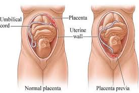
Vasa Previa occurs when the fetal blood vessel(s) from the placenta or umbilical cord cross the entrance to the birth canal, beneath the baby. Vasa previa can result in rapid fetal hemorrhage (occurs from the vessels tearing when the cervix dilates or membranes rupture) or lack of oxygen (if the vessels become pinched off as they are compressed between the baby and the walls of the birth canal). The aberrant vessels result from velamentous insertion of the cord, bilobed or succenturiate lobed placenta.

Symptoms

Vasa previa can be asymptomatic but can also present with sudden onset of abnormally heavy or small amounts of painless vaginal bleeding in the second or third trimester of pregnancy. Source of blood should always be investigated to determine whether the blood is maternal or fetal if the baby is not in distress.

Warning Signs

Vasa previa might be present if any of the following conditions exist: low-lying placenta (may be caused by previous miscarriages followed by curreting of the uterus (D&C) or uterine surgeries, which can cause scarring in the uterus), bilobed or succenturiate-lobed placentas, velamentous insertion of the cord, pregnancies resulting from in-vitro fertilization or multiple pregnancies. Vasa previa bleeding is painless. Other obstetrical or birthing bleeding complications are not necessarily painless.

http://vasaprevia.com/Vasa-Previa-Fact-Sheet

Vasa previa is an extremely rare but devastating condition in which fetal umbilical cord blood vessels cross or run in close proximity to the inner cervical os (the internal opening in the cervix separating the uterine cavity from the vagina). These vessels course within the membranes, unsupported by the umbilical cord or placental tissue, and are at risk of rupture if the supporting membranes are damaged. Vasa previa carries a high mortality rate—50percent of undiagnosed cases end in the death of the fetus. Fortunately, this condition is rare, occurring in only one out of every 2,000 pregnancies.

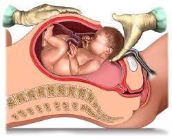
The normal umbilical cord begins right off the placenta, and it carries two arteries and one vein directly into the baby’s body. In vasa previa, the origin of the cord is in the membranes near the placenta, so there is an area in the membranes where the blood vessels are not in the cord at all. Even worse, this area of the membranes containing the cord blood vessels is in front of the internal cervical os. Without the protection of the tough, fibrous cord, the blood vessels have little support. When labor begins and the mother’s water breaks, the unsupported vessels in and around the umbilical cord can tear, resulting in the death of the baby from blood loss within two to three minutes unless an emergency. C-section is performed.

Also known as a Caesarean section, a C-section is when a mother delivers her baby through an incision made in her abdomen and uterus.

http://www.pregnancycorner.com/giving-birth/c-section.html

If you are having difficulty grasping what occurs in vasa previa, it may be helpful to understand the origin of the condition’s name, which is derived from Latin. Vasa is the plural form of the vas, which denotes a vessel (it’s the root of English words like vase), and previa (from which we also get previous) can be broken down into two components—pre, meaning before, and via, meaning way. Vasa previa can, therefore, be understood to mean roughly, “vessels in the way of the baby.”

For more on this

http://www.pregnancycorner.com/being-pregnant/complications/vasa-previa.html

http://vasapraevia.co.uk/

17q21.31 microdeletion syndrome is a condition caused by a small deletion of genetic material from chromosome 17. The deletion occurs at a location designated as q21.31. People with 17q21.31 microdeletion syndrome may have developmental delay, intellectual disability, seizures, hypotonia. distinctive facial features, and vision problems. Some affected individuals have heart defects, kidney problems, and skeletal anomalies such as foot deformities. Typically their disposition is described as cheerful, sociable, and cooperative. The exact size of the deletion varies among affected individuals, but it contains at least six genes. This deletion affects one of the two copies of chromosome 17 in each cell. The signs and symptoms of 17q21.31 microdeletion syndrome are probably related to the loss of one or more genes in this region.

Click on the colored link’s below for more help.

Advocacy and Support Organizations

Health Questions

Recommended Apps

Scientific Literature

Symptoms,

Diagnosis, and Treatment

Clinical Trial Information

2016

The chromosome 17q21.31 microdeletion syndrome has been renamed. The condition is now called the Koolen de Vries syndrome (KdVS; MIM #610443). The syndrome is caused by either the fact that a small part of chromosome 17 is missing (17q21.31 microdeletion) or a defect in a single gene: the KANSL1-gene.

http://17q21.com/en/publications.php

Koolen-de Vries syndrome 17q21.31 Microdeletion syndrome Questionnaire

http://www.designer-illusions.com/KdVS/QuestionnaireKdVS.pdf

Abstract

BACKGROUND:

Oesophageal atresia (OA) and mandibulofacial dysostosis (MFD) are two congenital malformations for which the molecular bases of syndromic forms are being identified at a rapid rate. In particular, the EFTUD2 gene encoding a protein of the spliceosome complex has been found mutated in patients with MFD and microcephaly (MIM610536). Until now, no syndrome featuring both MFD and OA has been clearly delineated.

RESULTS:

We report on 10 cases presenting with MFD, eight of whom had OA, either due to de novo 17q21.31 deletions encompassing EFTUD2 and neighboring genes or de novo heterozygous EFTUD2 loss-of-function mutations. No EFTUD2 deletions or mutations were found in a series of patients with isolated OA or isolated oculoauriculovertebral spectrum (OAVS).

CONCLUSIONS:

These data exclude a contiguous gene syndrome for the association of MFD and OA, broaden the spectrum of clinical features ascribed to EFTUD2 haploinsufficiency, define a novel syndromic OA entity, and emphasize the necessity of mRNA maturation through the spliceosome complex for global growth and within specific regions of the embryo during development. Importantly, the majority of patients reported here with EFTUD2 lesions were previously diagnosed with Feingold or CHARGE syndromes or presented with OAVS plus OA, highlighting the variability of expression and the wide range of differential diagnoses.

http://www.ncbi.nlm.nih.gov/pubmed/23188108

Clinical Trails Finder

Finding the right clinical trial for 17q21.31 Microdeletion Syndrome can be challenging. However, with Trials-Finder (which uses the Reg4ALL database and privacy controls by Private Access), you can permit researchers to let you know opportunities to consider – all without revealing your identity.

https://www.trialsfinder.org/?ga_id=GA9

What Can Unique Do To Help?

Having a child with a rare chromosome disorder can be a huge shock and can stir up a whole range of emotions and a great desire to learn more about your child’s disorder. Most of us who help run the group have been through these experiences and know how you are feeling. Most parents’ first reaction, quite understandably, is to “find” another, an older child with the same disorder as their child. While this might be possible for some, it still does not mean that the two children will develop in the same way. However, just talking to other parents with a child with a rare chromosome disorder can be a great relief and can help to alleviate feelings of isolation and “why me?”

As part of its services, Unique runs telephone (+44 (0) 1883 723356) and email (info@rarechromo.org) helplines for new and existing member families and professionals to find out more information about the group and about specific rare chromosome disorders. We have developed and maintain a comprehensive offline computerized database detailing the lifetime effects of specific rare chromosome disorders among our members. By Spring 2013 over 10,000 families will have joined our membership, representing more than 14,000 individuals with a rare chromosome disorder registered on our database, the vast majority being new cases never reported in published medical journals. New families are joining us daily. If you go to our registered disorders page on this website you will be able to see all the different chromosomal disorders, with their genotypes where known, occurring among our members. The offline database can be used to link families on the basis of a specific rare chromosome disorder. Often of more practical benefit, however, is to link families on the basis of problems as they arise, whether these are medical, developmental, behavioral, social, educational and so on. We also maintain close links with other similar groups around the world, thus increasing the “pool” of possible family contacts. Information about a specific rare chromosome disorder can be prepared from the Unique database while not revealing the identity of the families concerned.

http://www.rarechromo.org/html/WhatWeDoToHelp.asp

Fewer than 100 children in the UK are diagnosed each year with neuroblastoma. Most children who get this cancer are younger than five years old. Neuroblastoma is the second most common solid tumor in childhood, and it makes up 8% of the total number of children’s cancers.

More children than ever are surviving childhood cancer. There are new and better drugs and treatments. But it remains devastating to hear that your child has cancer, and at times it can feel overwhelming. There are many healthcare professionals and support organizations to help you through this difficult time.

Understanding more about the cancer your child has and the treatments that may be used can often help parents to cope. Your child’s specialist will give you more detailed information. If you have any questions it’s important to ask the specialist doctor or nurse who knows your child’s individual situation.

http://www.macmillan.org.uk/cancerinformation/cancertypes/childrenscancers/typesofchildrenscancers/neuroblastoma.aspx

Signs and symptoms of neuroblastoma

The signs and symptoms of neuroblastoma vary widely, depending on the size of the tumor, where it is, how far it has spread, and if the tumor cells secrete hormones.

Many of the signs and symptoms below are more likely to be caused by something other than neuroblastoma. Still, if your child has any of these symptoms, check with your doctor so the cause can be found and treated, if needed.

Signs or symptoms caused by the main tumor

Tumors in the abdomen (belly) or pelvis: One of the most common signs of a neuroblastoma is a large lump or swelling in the child’s abdomen. The child might not want to eat (which can lead to weight loss). If the child is old enough, he or she may complain of feeling full or having belly pain. But the lump itself is usually not painful to the touch.

Sometimes, a tumor in the abdomen or pelvis can affect other parts of the body. For example, tumors that press against or grow into the blood and lymph vessels in the abdomen or pelvis can stop fluids from getting back to the heart. This can sometimes lead to swelling in the legs and, in boys, the scrotum.

In some cases the pressure from a growing tumor can affect the child’s bladder or bowel, which can cause problems urinating or having bowel movements.

Tumors in the chest or neck: Tumors in the neck can often be seen or felt as a hard, painless lump.

If the tumor is in the chest, it might press on the superior vena cava (the large vein in the chest that returns blood from the head and neck to the heart). This can cause swelling in the face, neck, arms, and upper chest (sometimes with a bluish-red skin color). It can also cause headaches, dizziness, and a change in consciousness if it affects the brain. The tumor might also press on the throat or windpipe, which can cause coughing and trouble breathing or swallowing.

Neuroblastomas that press on certain nerves in the chest or neck can sometimes cause other symptoms, such as a drooping eyelid and a small pupil (the black area in the center of the eye). Pressure on other nerves near the spine might affect the child’s ability to feel or move their arms or legs.

Signs or symptoms caused by cancer spread to other parts of the body

About 2 out of 3 neuroblastomas have already spread to the lymph nodes or other parts of the body by the time they are found.

Lymph nodes are bean-sized collections of immune cells found throughout the body. Cancer that has spread to the lymph nodes can cause them to swell. These nodes can sometimes be felt as lumps under the skin, especially in the neck, above the collarbone, under the arm, or in the groin. Enlarged lymph nodes in children are much more likely to be a sign of infection than cancer, but they should be checked by a doctor.

Neuroblastoma often spreads to bones. A child who can talk may complain of bone pain. The pain may be so bad that the child limps or refuses to walk. If it spreads to the bones in the spine, tumors can press on the spinal cord and cause weakness, numbness, or paralysis in the arms or legs. Spread to the bones around the eyes is common and can lead to bruising around the eyes or cause an eyeball to stick out slightly. Cancer can also spread to other bones in the skull, causing bumps under the scalp.

If cancer spreads to the bone marrow (the inner part of certain bones that makes blood cells), the child may not have enough red blood cells, white blood cells, or blood platelets. These shortages of blood cells can result in tiredness, irritability, weakness, frequent infections, and excess bruising or bleeding from small cuts or scrapes.

A special widespread form of neuroblastoma (known as stage 4S) occurs only during the first few months of life. In this special form, the neuroblastoma has spread to the liver, to the skin, and/or to the bone marrow (in small amounts). Blue or purple bumps that look like small blueberries may be a sign of spread to the skin. The liver can become very large and can be felt as a mass on the right side of the belly. Sometimes it can grow large enough to push up on the lungs, which can make it hard for the child to breathe. Despite the fact that the cancer is already widespread when it is found, stage 4S neuroblastoma is very treatable, and often shrinks or goes away on its own. Almost all children with this form of neuroblastoma can be cured.

http://www.cancer.org/cancer/neuroblastoma/detailedguide/neuroblastoma-signs-and-symptoms

Neuroblastoma is a rare aggressive childhood cancer. About 100 children are diagnosed in the UK each year.

NBUK works exclusively for these children in raising funds for British research into the disease and offering information and support to families affected by neuroblastoma.

https://www.justgiving.com/NeuroblastomaUK