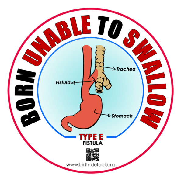
There are 5 types of (EA) Esophageal Atresia explained with video and logo’s

We have been helped by Surgeons to bring you this page

You can save these logo’s and use them yourselves

Videos have been added and your find some are Narrated by the Surgeons

Find at the Bottom – Tracheobronchomalacia (TBM)


There are 5 types of (EA) Esophageal Atresia explained with video and logo’s

Basic understanding of what Esophageal Atresia is, in the video on the right hand side.

Oesophageal atresia and tracheo-oesophageal fistula

The upper part of the oesophagus doesn’t connect with the lower oesophagus and stomach. It usually ends in a pouch, which means food can’t reach the stomach.  It often happens along with another birth defect called a tracheo-oesophageal fistula, which is a connection between the lower part of the oesophagus and the windpipe (trachea). OA-TEF and EA-TEF are one in the same condition depending on where you live in the world as to what you call it.

Australia and England this is known as:

Oesophageal atresia and tracheo-oesophageal fistula = OA-TOF

In some parts of the world, this is known as  Esophageal Atresia – EA – but what you see below is the same where ever you live.

Esophageal Atresia/Tracheo-Esophageal Fistula = EA-TEF


Type A

Thank You, Roze Diamond, for translating these into Spanish for us, and Karim Ocanas for checking the medical part.

Gross type A, Vogt type 2

Type A is when the upper and lower parts of the esophagus do not connect and have closed ends, like dead-end streets that don’t connect. In this type, no parts of the esophagus attach to the trachea, this is known as Pure Esophageal Atresia, with no TEF.

Tipo A.
Gross tipo A, Vogt tipo 2.
El tipo A es cuando las porciones superior e inferior (o proximal y distal) del esófago no están conectadas y tienen las puntas cerradas (o en fondo de saco ciego), igual a calles sin salida que no se conectan. En este tipo de atresia las porciones del esófago no se conectan a la tráquea, esto se conoce como Atresia Esofágica (AE) pura sin fístula.

Typ A

Występuje wtedy, gdy górna i dolna część przełyku nie łączą się i mają zamknięte końce, jak ślepe uliczki, które się nie łączą. W tym typie żadna część przełyku nie przyczepia się do tchawicy, jest to znane jako czysta atrezja przełyku, bez przetoki.

Type B

Gross type B, Vogt type 3A

Type B is very rare. In this type, the upper part of the esophagus is attached to the trachea, but the lower part of the esophagus has a closed-end. With this type of EA, food and saliva can travel directly into the lungs, before surgical correction is performed.

Tipo B.
Gross tipo B, Vogt tipo 3A.
El tipo B es muy raro. En este tipo la porción superior (o proximal) del esófago está conectada a la tráquea, pero la parte inferior (o distal) del esófago tiene la punta cerrada. Si no se lleva a cabo la corrección quirúrgica tanto la comida como la saliva pueden ir directamente a los pulmones.

Typ B

Wystepuje bardzo rzadko, w tym typie górna część przełku jest połączona z tchawicą ale dolna część przełyku jest zamknięta. W tym typie atrezji pokarm i ślina wędruje prosto do płuc przed wykonaniem korekcji chirurgicznej.

Stent to me by DR. Steven Rothenberg MD

Here is the video link for a long gap with proximal video.

Real-life Surgeons at work Just Click the link below REAL VIDEO

https://www.dropbox.com/s/1npoaksay2xf0z2/Long%20gap%20EA%20with%20prox%20fistula%20low%20res.mp4?dl=0&fbclid=IwAR04Ubbms-VEyP4bYmBkbCvB3ApfGzoKWxFnk7WUYPPXdChlnkWK6SSdo00


Type C

Gross type C, Vogt type 3B

Type C is the most common type. In this type, the upper part of the esophagus has a closed end and the lower part of the esophagus is attached to the trachea, with a distal TEF, or a TEF connecting between the lower pouch of the esophagus and the trachea.

Tipo C.
Gross tipo C, Vogt tipo 3B.
El tipo C es el más común. En este tipo la porción superior (o proximal) del esófago tiene la punta cerrada y la parte inferior del esófago está conectada a la tráquea, con una fistula traqueoesofágica (FTE) distal, o una FTE conectando entre la punta inferior (o distal) del esófago y la tráquea.

Typ C

Jest najczęstszym typem. W tym typie górna część przełyku jest zamknięta a dolna część jest połączona z tchawicą z dystalną przetoką pomiędzy dolną częścią przełyku a tchawicą

Stent to me by DR. Steven Rothenberg MD

Dr. Steve Rothenberg performs a thoracoscopic repair of a Type III Tracheoesophageal Fistula in a 2.6kg infant using the 3mm JustRight Sealer.

Here is the video link for a classic type C.

Type D

Gross type D, Vogt type 3C

Type D is the rarest and most severe. In this type, the upper and lower parts of the esophagus are not connected to each other, but each is connected separately to the trachea. Dual TEF, where there is both a TEF connecting between the upper pouch of the esophagus and the trachea and a TEF connecting the lower pouch of the esophagus and the trachea.

Tipo D.
Gross tipo D, Vogt tipo 3C.
El tipo D es la AEFTE doble, es la más rara y severa. En este tipo, las porciones superior e inferior del esófago no están conectadas una a la otra, pero cada una de las partes está conectada individualmente a la tráquea.
En la AEFTE existen ambos tipos de atresia: la bolsa superior del esófago conectada a la tráquea y la bolsa inferior del esófago también conectada a la tráquea.

Typ D

Jest najrzadszym i najcięższym. W tym typie górna i dolna część przełyku nie są ze sobą połączone, ale każda jest połączona osobno z tchawicą. Podwójna przetoka która łączy zarówno górną torebkę przełyku z tchawicą, jak i dolną torebkę przełyku z tchawicą


Type E Also known as H

Gross type E

TEF without esophageal atresia or so-called H-type fistula, H-Type TEF, where there is a TEF connecting between the esophagus and the trachea, but there is no EA. So this means that Children with an H-Type TEF, unlike the other types, are often diagnosed later in infancy or childhood. People with an H-type TEF can swallow, but often cough and choke during swallowing, especially with liquids, and may present with recurrent pneumonia. The exact prevalence of this form is not known (because they tend to be diagnosed late, but rarely as adults)

(this is known as Gross type E)

Gross tipo E.
La Fistula Traqueoesofágica sin atresia, o fistula tipo H, FTE Tipo H, es aquélla donde hay una FTE conectando entre el esófago y la tráquea, pero no hay AE. Esto significa que los niños con FTE Tipo H, a diferencia de los otros tipos, son diagnosticados tardíamente en su infancia o niñez. La gente con FTE Tipo H pueden toser frecuentemente y atragantarse mientras tragan, especialmente con líquidos. Además pueden presentar neumonías recurrentes. La prevalencia exacta de este tipo no es conocida (debido a que ellos tienden a ser diagnosticados tardiamente, pero raramente en la edad adulta.)

Typ E,

Przetoka bez atrezji przełyku lub tak zwanej przetoki typu H, gdzie istnieje przetoka łącząca przełyk z tchawicą, ale nie ma atrezji. Oznacza to, że dzieci z przetoką typu H, w przeciwieństwie do innych typów, często diagnozuje się później w wieku niemowlęcym lub dzieciństwie. Osoby z przetoką typu H mogą połykać, ale często kaszlą i duszą się podczas połykania, szczególnie płynów i może u nich występować nawracające zapalenie płuc. Dokładna częstość występowania tej postaci nie jest znana (ponieważ zwykle diagnozuje się ją późno, ale rzadko u dorosłych)

Stent to me by DR. Steven Rothenberg MD

Here is the video Link for H type TEF. Click on this link below

Real-life Surgeons at work Real Video.

https://www.dropbox.com/s/f38jdzsfz97qeeb/H%20type%20TEF%20IPEG%20Video.mov?dl=0&fbclid=IwAR1k4D0pgZt3sh7Zod_09R586mMas5jkMbSdwzuqo3qa1tcIwsZfh0cTpBA

There are 5 types of (EA) Esophageal Atresia explained with video and logo’s

Watch this video, where you see the Parents talking about the fantastic work Steven Rothenburg and his team did on their child.

Endoscopy of H-Type Tracheoesophageal fistula

Romaine Johnson

Tracheoesophageal Fistula (TEF, TOF)

Meet Dr. Rajan Garg Pediatric Surgeon (AllMS)

This Surgeon is in India and has shown support for this website over the past few years.

This is in Hindi This young Surgeon is talking about doing the first thoracoscopic TEF Repair in India.

Might apper after a repair of EA/TEF

In some patients, TM/TBM becomes apparent soon after the repair of EA/TEF when they cannot be extubated.

Tracheobronchomalacia (TBM) is a rare disorder characterized by the collapse of the trachea (windpipe) and main bronchi (main airways) due to the weakness in the cartilaginous support structures. Tracheobronchomalacia is a clinically challenging condition, frequently undiagnosed or misdiagnosed in pediatrics, and includes many airway pathologies that cause either fixed or dynamic airway narrowing. Symptoms include cough, shortness of breath, persistent wheezing, inability to clear secretions and fatigue during exercise. Symptoms can be nonspecific and frequently overlap with other more common respiratory conditions. This is the main reason for the delay in diagnosis. Cough is a common symptom of TBM. Patients with TBM may have a characteristic barking seal cough.

Clinical presentation of TM/TBM includes a range of non-specific respiratory symptoms, depending on the location, extent, and severity of the airway collapse. Many children with TM/TBM do not show symptoms until age 2–3 months. However, in cases with long-segment TM/TBM, symptoms may begin at birth.

There is a Group for this. Click on the link here to go to the page on this website.

Born Unable to Swallow Parent run Support Groups

Interview with Todd Ponsky MD, Assistant Editor Journal of Pediatric Surgery on Posterior tracheopexy for severe tracheomalacia

https://www.youtube.com/watch?v=bIpIYoH3HLg

.