Respect Earned , Respect Given

I have used this photo above because History plays a big part in our lives

What you find on this page will be info I have come across over the time doing this site,

that I have felt most wouldn’t know,

but I found very interesting because most of us never think about how or where

these ideas have come from.

I hope you like what you read on this page

What you read on this page is how far the Doctors have come. The history of oesophageal atresia and tracheo-oesophageal fistula

The history of oesophageal atresia commenced in the year 1670 with Durston’s description of “A narrative of a monstrous birth at Plymouth”. However, the most significant contribution in the 17th century was made by Gibson, who clearly described the clinical picture and necropsy findings in a baby with a proximal oesophageal atresia and a distal tracheo-oesophageal fistula. The 18th century was singularly lacking in contributions to the literature, but there were many important presentations during the 19th century. Early in that century, Martin in France and Hill in the United States of America contributed to the literature; subsequently, many anecdotal references can be found particularly in England, and ultimately a surgical attempt to correct the anomaly was made in 1888 by Steele in London. With this, the pre-surgical era gave way to the surgical era, and at a later stage pre-survival era was to give way to the survival phase. Reference will be made in the text to these various phases in the history and to the many pioneering surgical endeavours which took place in the earlier years of the 20th century. It will be seen that the surgical efforts employed palliative procedures, indirect attacks on the problem and finally direct attacks.

The classic successes by indirect methods which were achieved by Ladd and Leven in babies born in 1939 were to be followed by the first successful end-to-end anastomosis which was achieved in Ann Arbor, Michigan, by Cameron Haight. Others had attempted primary anastomosis, and Lanman, in particular, deserves mention. However, it was Cameron Haight who opened a new era in the history of the treatment of oesophageal atresia; the recent history is largely concerned with increasing experience and refinements in technique. For the future historian, many pathways can still be explored, including oesophageal replacement, the unstable trachea, follow-up studies and research projects. This history also included comments on the history of the less frequently encountered anomalies and such aspects as classification and radiology. The history of oesophageal atresia and tracheo-oesophageal fistula is a mini history of surgery – “oesophageal atresia is the epitome of modern surgery”.

Please watch this video to learn how the TPN method was BORN. If it wasn’t for the drive to find a way to keep our Children alive by finding a way of feeding through the vain then your child or mine would not be here today, I for one am very grateful he never gave up, reason for me my own Daughter was the First in the world when she was young to be feed by TPN

Why the first you may ask, it took 11 world consultants, 3 were in the USA to work out how this could be done on a child with a very rare metabolic condition, and they cracked it. But if it wasn’t for this Dr below then god only knows what the outcome would have been. I went looking to find out who thought of TPN, this is what I found.

If you wont to know how it came about then watch the video I have below.

Stanley Dudrick, MD, FACS, Professor of Surgery at The Commonwealth Medical College (TCMC) was honored as a “Living Legend” by The International Society of Small Bowel Transplantation on June 12, 2015 at its annual conference in Buenos Aries, Argentina. Known as “the father of intravenous feeding,” Dr. Dudrick pioneered total parenteral nutrition (TPN) in the early 1960’s. Originally from Nanticoke, PA, he was the first physician to successfully feed people through an I.V., totally bypassing the normal digestive system. Prior to TPN, patients who could not eat or digest food routinely died of starvation. Today, more than 10 million premature babies and 20 million adults owe their lives to Dr. Dudrick’s brilliant invention.

The International Society of Small Bowel Transplantation annually selects recipients as Living Legends to commemorate distinguished careers that have impacted health care through notable contributions to transplantation, research, and education.

Dr. Dudrick explained, “TPN is a lifesaver for patients with short bowel syndrome. They have to be fed by vein or else they die. Some of these patients have a bowel transplant. TPN keeps the patients alive while they’re waiting for transplants. Then TPN will feed the patient until the new bowel sustains the patient, and it serves as a safety net in case the transplant doesn’t work.”

Dr. Dudrick has received numerous accolades for his work. In Skawina, Poland, the Stanley Dudrick, MD Memorial Hospital stands in his honor. Last summer, Dr. Dudrick was named a “Hero of American Surgery” by the American College of Surgeons.

William F. Iobst, MD, Vice Dean and Vice President of Academic Affairs at TCMC, said, “Dr. Dudrick deserves the title ‘living legend.’ TPN is considered one of the three most important medical advances of the last century. We are incredibly proud to have him on the faculty at TCMC, and fortunate he has returned to his home community.”

I asked if I could write this and use this video on this site.

As most of you might know my own daughter is tube fed, but this got me thinking who invented this, a year ago this was posted on a Surgeons facebook page I am friends with, the Birth of Tube Feeding.

The Post Read.

What a GREAT video on the history of how my father Jeffrey Ponsky invented the PEG feeding tube now used by billions around the world. He is one the most humble people I have known so he rarely tells the whole story.

My greatest hero, always around as a father while he was revolutionizing medicine and inventing PEG. He was recently honored with an appointment as the Lynda and Martin Younker Professor of Developmental Endoscopy and tells the story here in his video.

I love you dad and hope that I can one day accomplish a fraction of what you have accomplished and be a fraction of the father you are.

(I found this very interesting)

What a GREAT video on the history of how my father Jeffrey Ponsky invented the PEG feeding tube now used by billions around the world. He is one the most humble people I have known so he rarely tells the whole story. My greatest hero, always around as a father while he was revolutionizing medicine and inventing PEG. He was recently honored with an appointment as the Lynda and Martin Younker Professor of Developmental Endoscopy and tells the story here in his video. I love you dad and hope that I can one day accomplish a fraction of what you have accomplished and be a fraction of the father you are.

Δημοσιεύτηκε από Todd Ponsky στις Τετάρτη, 26 Ιουλίου 2017

The First thoracoscopic TEF was then carried out in Denver USA again by Steven Rotherburg details I have found, 2000

Thoracoscopic repair of esophageal atresia and tracheo­esophageal fistula in neonates: the current state of the art.

Rocky Mountain Hospital for Children, Denver, CO, USA

Abstract
The first thoracoscopic esophageal atresia with tracheo-esophageal fistula
(EATEF) repair was performed in March of 2000. This report evaluates the
results and evolution of the technique over the last decade. Thoracoscopic
esophageal atresia repair has proven to be an effective and safe technique.
Initial experience resulted in a higher stricture rate but this improved with
experience and changes in technique over the last decade. The outcomes are
similar to or superior to that of an open thoracotomy and avoid the
musculoskeletal morbidity associated with that technique.

Keywords
Tracheo-esophageal fistula
Esophageal atresia
Thoracoscopy
Introduction
Esophageal atresia (EA) with or without a tracheo-esophageal (TEF) fistula is
one of the rarer congenital anomalies occurring in one in 3,000 births.
Traditionally these patients have presented shortly after birth because of an
inability to pass an oro-gastric tube, respiratory distress, or an inability to

tolerate feedings. The condition maybe associated with other major congenital
anomalies (VATER syndrome), or may be an isolated defect [ 1 – 3 ].
Improvements in maternal-fetal ultrasound have resulted in prenatal diagnosis
in a number of cases. This allows the surgeon to plan for delivery and eventual
surgery. Patients with a tracheo-esophageal fistula require relatively emergent
surgical intervention to prevent aspiration of gastric acid and over distension
of the intestines. Those with pure atresia can be dealt with in a more leisurely
fashion as long as the infants’ oral secretions are controlled by continuous or
intermittent suction.
Advancements in technique and instrumentation in pediatric endoscopic
surgery have allowed significantly more complex and delicate procedures to be
performed, even in small neonates. Over the last 20 years, the number and
breadth of minimally invasive surgical (MIS) procedures performed in infants
have increased dramatically including the repair of esophageal atresia. [ 4 – 8 ].
In 1999, a stepping stone was laid when a successful thoracoscopic repair of a
pure esophageal atresia was completed in a 2-month-old male [ 9 ]. In 2000, we
reported on the first successful repair of an esophageal atresia with tracheoesophageal
fistula (EATEF) in a newborn using a completely thoracoscopic
approach [ 10 ] and 2 years later reported on the first significant series [ 11 ].
These milestones allowed for a more widespread adoption of these techniques
so that numerous pediatric surgical units around the world are now performing
minimally invasive EATEF repair. There are now reports from over the world
reporting excellent results using a thoracoscopic approach for this disease.
This paper will review the development and technical advances of this
technique as well as review the author’s personal experience.

Technique
The technique has been previously well described with mild variations between
centers. A brief description will be given here for clarity. General endotracheal
anesthesia is administered and an attempt is made to maintain low peak
pressures until the fistula is ligated to prevent over distension of the abdomen.
Single lung ventilation is not attempted, instead a low flow, low pressure of
CO (4 mmHg, 1 L/min) is used to collapse the right lung and create space.

Positioning

Once the endotracheal tube is secured, the patient is placed in a modified
prone position with the right side elevated approximately 30º. If there is a
right-sided arch then a left sided approach is used. This positioning gives the
surgeon access to the area between the anterior and posterior axillary line for
trocar placement, while allowing gravity to retract the lung away from the
posterior mediastinum. This arrangement gives excellent exposure of the
fistula and esophageal segments without the need of an extra trocar for a lung
retractor. The surgeon and the assistant stand in front of the patient and the
monitor is placed behind the patient. This allows the surgeon and the assistant
to work in line with the camera towards the point of dissection. The assistant
should not be placed on the opposite side of the table as this will place him at
a complete paradox with the telescope. The scrub nurse can be on either side
of the patient depending on the room layout. Because of the fine manipulation
necessary the surgeon and the assistant should position themselves so that they
are in the most ergonomic and comfortable position.
AQ2
Port placement
Port placement is extremely important because of the small chest cavity and
the intricate nature of the dissection and reconstruction. The procedure can be
performed with three ports but occasionally a fourth port is necessary to retract
the lung.
The initial port (3–5 mm) is placed in the fifth intercostal space posterior to
the tip of the scapula. This is the camera port and gives the surgeon excellent
visualization of the posterior mediastinum in the area of the fistula and
eventual anastomosis. A 30º lens is used to allow the surgeon to “look down”
on his instruments and avoid “instrument dueling”.
The two instrument ports are placed to achieve a ninety degree angle at the
presumed site of the anastomosis. The first port is placed in the mid-axillary
line one to 2 interspaces above the camera port. This upper port is 5 mm to
allow for the introduction of a clip applier and suture. A 3 mm port can be
used if the surgeon wishes to suture ligate the fistula and passes the sutures
through the chest wall. The lower port is 3 mm in size and is placed one or
two intercostal spaces below, and slightly posterior to the camera port

(Fig. 1 ). Ideally the instrument tips will approximate a right angle (90º) at the
level of the fistula. This positioning will facilitate suturing the anastomosis. A
fourth port can be placed either higher or lower in the thoracic cavity to help
retract the lung, but this has not been necessary in the majority of cases. The
operation then follows the same pattern as for the open procedure.
Fig. 1
Trocar placement with the infant in a modified prone position with the left side
elevated 30º. The 30º scope is placed in the port just below and posterior to the
tip of the scapula

Cont from report

Ligating the fistula
Once the chest has been insufflated and the lung collapsed, the surgeon must
identify the fistula. In most patients, the fistula is attached to the membranous
portion of the trachea just above the carina. This level is usually demarcated
by the azygos vein. After the azygos is identified, it should be mobilized for a short segment using
a curved dissector or scissors. The vein is then cauterized and divided. Some
advocate leaving the azygos intact, saying it improves vascularity of the area
and decreases the leak rate. This point is still debatable but the vein may be
preserved if desired.
With the vein divided, the lower esophageal segment is identified and followed
proximally to the fistula. Because of the magnification afforded by the
thoracoscopic approach it is easy to visualize exactly where the fistula enters
the back wall of the trachea. A 5 mm endo clip can then be applied safely or
the fistula can be suture ligated. Whichever technique is used, care should be
taken to avoid the vagus nerve (Fig. 2 ).
Fig. 2
Exposure of the fistula for ligation

Mobilizing the upper pouch
Attention is now turned to the thoracic inlet. The anesthesiologist places
pressure on the NG tube to help identify the upper pouch. The pleura overlying
the pouch are incised sharply and the pouch is mobilized with blunt and sharp
dissection. The plane between the esophagus and trachea can be seen well and
the two should be separated by sharp dissection. Mobilization of the upper
pouch is carried on as far as necessary up into the thoracic inlet or neck
depending on the length of the gap (Fig. 3 ). Fig. 3
Mobilization of upper pouch
Once adequate mobilization is achieved, the distal tip of the pouch is resected.
This should be an adequate section so that there is a sufficient opening to
prevent later stricture formation.
The anastomosis
With the two ends mobilized the anastomosis is performed using a 4-0 or 5-0
mono-filament absorbable suture in a single interrupted fashion. The back wall
is placed first and then an Ng tube is passed under direct vision into the lower
pouch and on into the stomach. The anterior wall is completed with the Ng
tube acting as a guide to prevent incorporation of the posterior wall and
ensuring patency of the anastomosis (Fig. 4 ).
Fig. 4
The completed anastomosis
8/27/2014 e.Proofing

Once the anastomosis is completed, a chest tube is placed through the lower
trocar site and the tip is placed near the anastomosis (under direct vision with
the endoscope). The other ports are removed and the sites are closed with
absorbable suture (Fig. 5 ).
Fig. 5
Trocar skin incisions at the end of the procedure. A chest drain is placed in the
left hand port

Author’s experience
From March 2000 to September 2012, 61 consecutive patients, 52 with
esophageal atresia and a distal tracheo-esophageal fistula and 9 with pure
esophageal atresia were referred to the author for repair. 16 had been
diagnosed pre-natally and were delivered at the high risk, perinatal/neonatal
center. Gestational age of the patients ranged from 30 to 40 weeks at the time
of delivery. Three other infants with EATEF were also referred to the author
during this period but were excluded because of size and associated anomalies.
These three weighed 800, 1,100 g with a Tetralogy of Fallot, and 1,800 g with
an omphalocele. In the thoracoscopic group, patients ranged in weight from
1.2 to 3.8 kg. Pre-operative evaluations revealed congenital heart disease in
22, including one patient with a Tetralogy of Fallot as well as a right-sided
aortic arch. One patient had a double aortic arch that was right-side dominant.
This patient underwent division of his left arch and repair of the EATEF
during the same procedure. Three patients had a high imperforate anus and one
had a cloaca. Six patients required intubation prior to surgery for increasing
respiratory distress.
The gap length was estimated pre-operatively based on the position of the tip
8/27/2014 e.Proofing

of the NG tube and the apparent bifurcation of the trachea as seen on the CXR.
This ranged from 2 to 4 1/2 vertebral bodies. At the time of surgery, the
longest gap in a patient with EATEF was closer to 4 1/2 vertebral bodies as
this patient had a trifurcation type fistula. The longest gap in a patient with
pure atresia was 7 vertebral bodies.
Results
Sixty of 61 procedures were completed successfully thoracoscopically. The
average operative time was 85 min (range 55–120 min). Esophageal contrast
studies were obtained on postoperative day (POD)# 3 in 1 patient, POD#4 in
30, and POD# 5 in 28 patients and the anastomosis was patent with no
evidence of a leak in 58/59. The first patient repaired with this technique had
clinical evidence of a leak, saliva in the chest tube, on POD# 4. He was kept
NPO and drainage stopped after 24 h. He was studied on day 8 with no
evidence of a leak. Two patients with a long gap pure atresia also had clinical
evidence of a small leak, which had sealed on a repeat study on day 9, and 10,
respectively. The one conversion was performed in a patient with an
unrecognized distal congenital esophageal stenosis. The anastomosis was
successfully completed thoracoscopically but because the NG tube could not
be passed into the stomach, the case was converted to ensure there was not a
false passage.
Discussion
Many recent studies have documented the long-term outcome of patients with
EA and TEF and the overall morbidity is significant, some related to the initial
surgical approach and technique [ 12 , 13 ]. The benefits of performing EA
repair using minimally invasive techniques are obvious but the technical
hurdles are significant. The greatest advantage is avoiding a postero-lateral
thoracotomy in a neonate. This has been shown to be associated with a high
degree of scoliosis and shoulder girdle weakness later in development
[ 14 – 16 ]. A more recent study by Lawal et al. comparing children who
underwent a thoracotomy vs. a thoracoscopic approach, showed a very high
rate of scoliosis (54 %) vs. 10 % in the thoracoscopy group. They also did
detailed measurements looking for chest wall asymmetry and found a much
higher rate in the thoracotomy group. And lastly, they polled the families

(There is a lot more on this click link below see photos etc)

https://rockymountainkidssurgery.com/util/forms/Thoracoscopic-Repair-of-Esophageal-Atresia.pdf

A 2-YEAR-OLD girl born without a windpipe now has a new one grown from her own stem cells.

This is How far it has come.

A 2-YEAR-OLD girl born without a windpipe now has a new one grown from her own stem cells, the youngest patient in the world to benefit from the experimental treatment.

Hannah Warren has been unable to breathe, eat, drink or swallow on her own since she was born in South Korea in 2010. Until the operation at a United States hospital, she had spent her entire life in a hospital in Seoul. Doctors there told her parents there was no hope and they expected her to die.

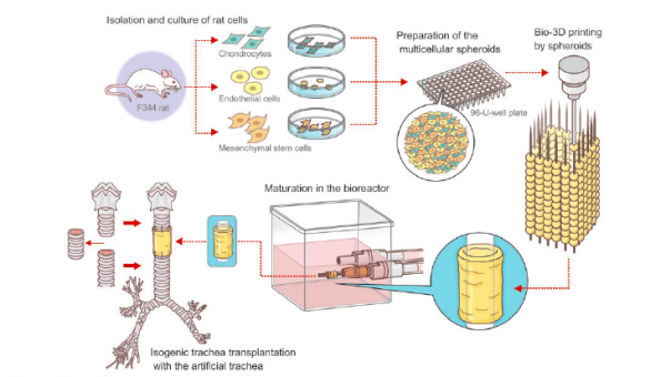
The stem cells came from Hannah’s bone marrow, extracted with a special needle inserted into her hip bone. They were seeded in a lab onto a plastic scaffold, where it took less than a week for them to multiply and create a new windpipe.

About the size of an 8cm tube of penne pasta, it was implanted on April 9 in a nine-hour procedure.

Early signs indicate the windpipe is working, Hannah’s doctors announced Tuesday, although she is still on a ventilator. They believe she will eventually be able to live at home and lead a normal life.

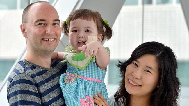
“We feel like she’s reborn,” said Hannah’s father, Darryl Warren.

“They hope that she can do everything that a normal child can do but it’s going to take time. This is a brand new road that all of us are on,” he said in a telephone interview. “This is her only chance but she’s got a fantastic one and an unbelievable one.”

Only about one in 50,000 children worldwide are born with the same defect. The stem-cell technique has been used to make other body parts besides windpipes and holds promise for treating other birth defects and childhood diseases, her doctors said.

The operation brought together an Italian surgeon based in Sweden who pioneered the technique, a pediatric surgeon at Children’s Hospital of Illinois in Peoria who met Hannah’s family while on a business trip to South Korea, and Hannah – born to a Newfoundland man and Korean woman who married after he moved to that country to teach English.

Hannah’s parents had read about Dr. Paolo Macchiarini’s success using stem-cell based tracheas but couldn’t afford to pay for the operation at his center, the Karolinska Institute in Stockholm. So Dr. Mark Holterman helped the family arrange to have the procedure at his Peoria hospital, bringing in Macchiarini to lead the operation. Children’s Hospital waived the cost, likely hundreds of thousands of dollars, Holterman said.


Part of OSF Saint Francis Medical Center, the Roman Catholic hospital considers the operation part of their mission to provide charity care, but also views it as a way to champion a type of stem-cell therapy that doesn’t involve human embryos, the surgeons said. The Catholic church opposes using stem cells derived from human embryos in research or treatment.

He said only one patient died, a 30-year-old man from Abingdon, Maryland, who had the operation in November 2011 to treat late-stage cancer of the windpipe. He died in March 2012 of uncertain causes, Macchiarini said.

Similar methods have been used to grow bladders, urethras and last year a girl in Sweden got a lab-made vein using her own stem cells and a cadaver vein.

Scientists hope to eventually use the method to create solid organs, including kidneys and livers, said Dr. Anthony Atala, director of Wake Forest University’s Institute for Regenerative Medicine. He said the operation on Hannah Warren “is really showing that the technique is workable.”

Hannah had breathing difficulties at birth and Korean doctors soon discovered the missing windpipe. They reconfigured her esophagus so that a breathing tube could go down it from her mouth to her lungs. The esophagus normally runs behind the windpipe and carries food to the stomach.

Korean doctors said she couldn’t live long with the tube and told her parents there was nothing more they could do.

Hannah outlived their expectations and has thrived despite the grim prognosis and other abnormalities including an undeveloped voice box that prevented her from speaking. Now that she has a windpipe and can breathe more normally, doctors expect the larynx to grow and function normally. She will work with speech therapists to help her learn to talk.

Dr. Holterman said Hannah will likely need a new windpipe in about five years, as she grows.

She breathes with help from a ventilator but no longer has a tube in her mouth that she’d lived with since shortly after birth, Dr Holterman said. She’s not yet able to eat normally, but doctors let her have her first taste ever of food – a few licks on a lollipop. Her father said she already has a discriminating taste and prefers chocolate Korean lollipops to the American kind.

“I asked her, ‘Is it good,’” he said, “and she immediately nodded her head.”

https://www.news.com.au/lifestyle/girl-2-gets-windpipe-made-from-stem-cells/news-story/a29827e262c68bf6569af8edc49cbd18

Mid 2017

The Regenova 3D printer placed spheroids in a 9 by 9 needle array in what can be described as “the Kenzan method.” Overall, 384 spheroids generated the 3D tubular trachea structure. The printed spheroids were then covered in with chondrocyte growth medium and endothelial cell growths prior to transplantation.

Testing and transplanting the trachea

The tensile strength of three of the 3D printed artificial tracheas was tested to failure using a DMT Tissue Puller. The test showed that the tracheas were strong enough to transplant into nine F344 rats.

The tracheas were surgically placed into the rats together with stents. The F344 rats were subsequent carefully monitored over 23 days and their activity noted.

According to the paper’s conclusions, the technology demonstrated the possibility of 3D printing further tracheal grafts, for use in trachea regeneration.

Japan researchers use Regenova bioprinter to 3D print windpipe without scaffolds

1950s

” Atresia of the oesophagus, with or without
tracheo-oesophageal fistula, has been a baffling
problem for the surgeon. If there is any surgeon
who has attempted to save the lives of patients
suffering from these malformations who has not
had many disappointments and numerous trials and
tribulations, I have not heard of him.”-W. E.
LADD (1944).
In this paper the clinical features, complications
of operation, and results of treatment are considered
in a personal series of 36 babies suffering
from congenital abnormalities of the oesophagus.
The patients were treated in the Children’s
Hospital, Birmingham, during the period June,
1953, to June, 1957. During this time, 44 such
babies were admitted and, as an indication of the
relative occurrence of these abnormalities, it may
be stated that the average annual admissions for
congenital hypertrophic pyloric stenosis were 85.
The classification of oesophageal abnormalities
used is that described by Gross in 1953 (Fig. 1).
Thirty-three babies belonged to Group C and one
each to Groups A, D, and E.
In 1955, Roberts, Carre, and Inglis noted that
Group C could be divided into two subgroups
(Fig. 2), namely, Group C(i) in which the fistula
enters the bifurcation of the trachea or the
bronchus and there is a considerable length of
atresia; these cases are unfavourable for primary
anastomosis (18 patients); Group C(ii) in which
the fistula enters the trachea high up and the
upper oesophageal segment overlaps the fistula
or the gap between the segments is not great (14
patients).
In one baby in the series, there were two fistulae
between the lower oesophagus and the trachea, one
being at the thoracic inlet and the other at the
bifurcation of the trachea. No similar case has
been described in the literature and this one has
provisionally been classified in both categories as
C(i)-(ii).
* Based on a paper read before the Society of Thoracic Surgeons
of Great Britain and Ireland on November 22. 1957.

A feature of the condition which has been neglected
is the association of atresia with maternal
hydramnios. It has long been believed that
atresias of the alimentary tract may cause hydramnios,
as the foetus is unable to swallow and absorb
amniotic fluid, and the incidence is particularly
marked in the case of oesophageal atresia
(Ballantyne, 1904; Scheurer, 1928; Brigham,
1929; Meyer, 1929). In the present series of 36
babies, 35 had oesophageal atresia and hydramnios
had been present in 13; the incidence may well be
higher than this, as details of the pregnancy have
not always been available.
CLINICAL SYMPTOMS AND SIGNS
The essential feature of total oesophageal
obstruction in the newborn is the inability of the
baby to swallow its saliva, so that a characteristic
fine frothy mucus is continually produced in the
mouth, unlike the rather coarse bubbles which are
frequently found for a time after birth (Belsey
and Donnison, 1950). The froth may be bilestained
owing to regurgitation of alimentary contents
through a tracheo-oesophageal fistula into the
trachea. Lecutier (1955) has given an interesting
account of ” paradoxical haematemesis ” in a baby
with a Group C abnormality, and one of the babies
in the present series was said to have ” vomited”
blood shortly after delivery.
Signs of respiratory obstruction with stridor may
be due to a fold of mucous membrane in the
trachea at the site of the fistula (Franklin and
Graham, 1953). Stridor was noted before operation
in only one baby in the series and recurred
after operation, but no tracheal fold was found at
post-mortem examination to account for it. If
feeds are attempted, ” spill-over” from the
obstructed oesophagus into the larynx causes
cough and cyanosis.